Copyright
©2012 Baishideng Publishing Group Co.
World J Gastroenterol. Feb 14, 2012; 18(6): 583-586
Published online Feb 14, 2012. doi: 10.3748/wjg.v18.i6.583
Published online Feb 14, 2012. doi: 10.3748/wjg.v18.i6.583
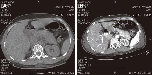
Figure 1 A computed tomographic scan of the abdomen revealed pancreatitis.
A: Plain computed tomographic (CT) scan; B: Enhanced CT scan.
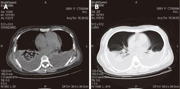
Figure 2 A computed tomographic scan of the chest revealed both sides pleural effusion, lung infection and pulmonary hypertension.
A: Mediastinal window; B: Lung window.
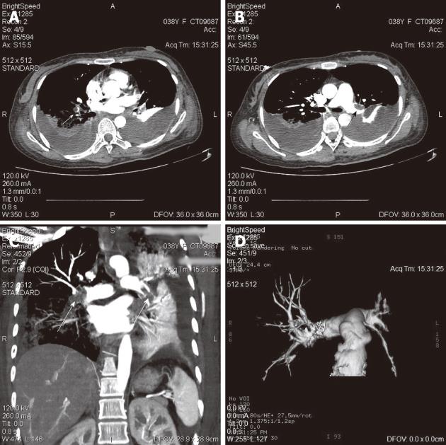
Figure 3 A Computer Tomography angiography of chest revealed pulmonary embolism (both down pulmonary arteries, left pulmonary artery and branch of right pulmonary artery).
A: Embolism of rihgt down pulmonary artery (arrow); B:Embolism of left down pulmonary artery (arrow); C:Embolism of left pulmonary artery and branch of right pulmonary artery in coronal view of chest-3D slab image (arrows); D: 3D reconstruction of pulmonary arteries.
- Citation: Zhang Q, Zhang QX, Tan XP, Wang WZ, He CH, Xu L, Huang XX. Pulmonary embolism with acute pancreatitis: A case report and literature review. World J Gastroenterol 2012; 18(6): 583-586
- URL: https://www.wjgnet.com/1007-9327/full/v18/i6/583.htm
- DOI: https://dx.doi.org/10.3748/wjg.v18.i6.583


