Copyright
©2012 Baishideng Publishing Group Co.
World J Gastroenterol. Dec 28, 2012; 18(48): 7405-7408
Published online Dec 28, 2012. doi: 10.3748/wjg.v18.i48.7405
Published online Dec 28, 2012. doi: 10.3748/wjg.v18.i48.7405
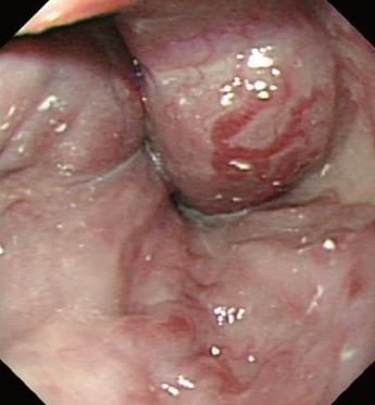
Figure 1 Gastroscopy showing severe esophageal and gastric varices.
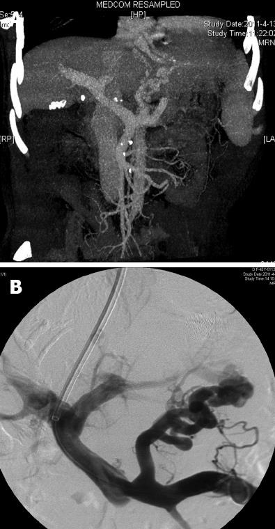
Figure 2 Portal venous-phase computed tomography and portal venography.
A: Portal venous-phase computed tomography showing classical appearance of portal hypertension with liver cirrhosis and splenomegaly; B: Portal venography showing the anatomy and direction of portal flow.
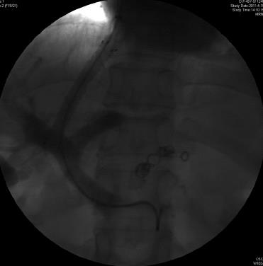
Figure 3 Completion venography of transjugular intrahepatic portosystemic shunt showing good flow and no contrast extravasation.
- Citation: Liu K, Fan XX, Wang XL, Wu XJ. Delayed liver laceration following transjugular intrahepatic portosystemic shunt for portal hypertension. World J Gastroenterol 2012; 18(48): 7405-7408
- URL: https://www.wjgnet.com/1007-9327/full/v18/i48/7405.htm
- DOI: https://dx.doi.org/10.3748/wjg.v18.i48.7405


