Copyright
©2012 Baishideng Publishing Group Co.
World J Gastroenterol. Dec 28, 2012; 18(48): 7290-7295
Published online Dec 28, 2012. doi: 10.3748/wjg.v18.i48.7290
Published online Dec 28, 2012. doi: 10.3748/wjg.v18.i48.7290
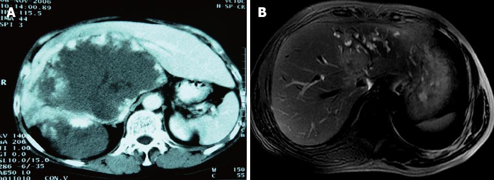
Figure 1 Computed tomography and magnetic resonance imaging of tumor.
A: Preoperative computed tomography scan (case 1) demonstrated a large hemangioma located in S1, S4, S5, S6, S7 and S8 of liver, and inferior vena cava (IVC) was involved and compressed severely (IVC circumference 190°, longitude 5 cm); B: Magnetic resonance imaging (case 3) showed the tumor located in S1, S5, S7 and S8, with a diameter of 5 cm; IVC (circumference 60°, longitude 2 cm), right hepatic vein thrombus, main hepatic vein as well as right portal vein were infiltrated by the tumor.
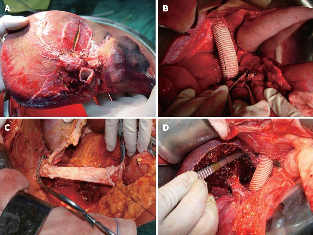
Figure 2 Main technical aspects of ex-situ liver surgery without veno-venous bypass.
A: Bench hepatectomy-cutting line of removed liver (case 3); B: Infiltrated inferior vena cava was replaced with a 20-mm ringed polytetrafluoroethylene graft; C: Reconstruction of inferior vena cava when polytetrafluoroethylene graft was removed (case 1); D: Anastomosis was performed in excised portal vein, hepatic artery and hepatic duct of residual right hepatic lobe (case 2).
-
Citation: Zhang KM, Hu XW, Dong JH, Hong ZX, Wang ZH, Li GH, Qi RZ, Duan WD, Zhang SG.
Ex-situ liver surgery without veno-venous bypass. World J Gastroenterol 2012; 18(48): 7290-7295 - URL: https://www.wjgnet.com/1007-9327/full/v18/i48/7290.htm
- DOI: https://dx.doi.org/10.3748/wjg.v18.i48.7290


