Copyright
©2012 Baishideng Publishing Group Co.
World J Gastroenterol. Dec 28, 2012; 18(48): 7166-7174
Published online Dec 28, 2012. doi: 10.3748/wjg.v18.i48.7166
Published online Dec 28, 2012. doi: 10.3748/wjg.v18.i48.7166
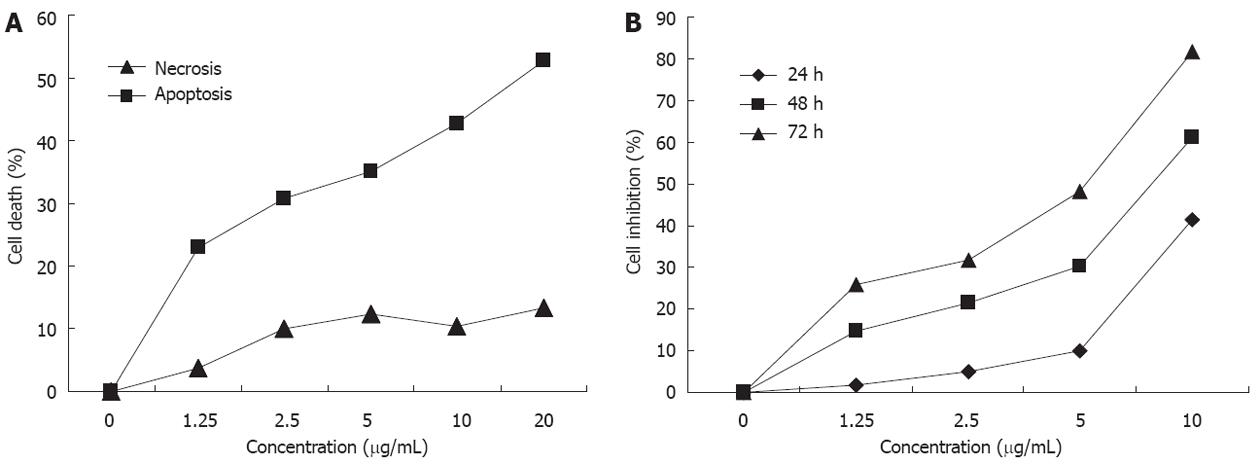
Figure 1 Inhibition of growth and lactate dehydrogenase release assay of HGC-27 cells after treatment with different concentrations of oridonin.
A: Inhibition of growth HGC-27 cells; B: Lactate dehydrogenase (LDH) release assay of HGC-27 cells. The change in the release of LDH caused by apoptosis was significant.
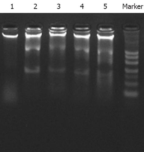
Figure 2 DNA ladder diagram after treatment with different concentrations of oridonin for 24 h.
1: Control; 2: 1.25 μg/mL oridonin; 3: 2.5 μg/mL oridonin; 4: 5 μg/mL oridonin; 5: 10 μg/mL oridonin.
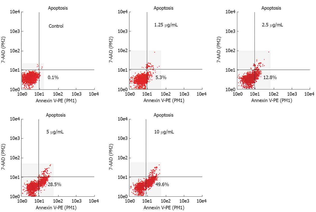
Figure 3 Analysis of apoptosis in HGC-27 cells.
Flow cytometric analysis showed that oridonin induced the apoptosis of HGC-27 cells in a dose dependent manner. The x-axis indicates the Annexin V-positive populations and the y-axis indicates the 7-AAD-positive populations. The lower right was the early apoptotic cells.
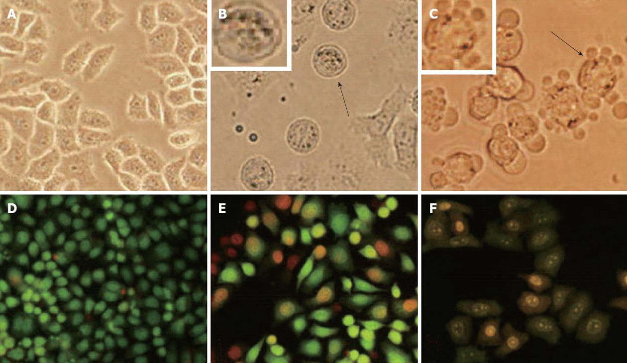
Figure 4 Morphological changes in HGC-27 cells after treatment with 10 μg/mL oridonin for 24 h and 48 h.
A: Negative; B: Treatment for 24 h; C: Treatment for 48 h; D: Acridine orange/ethidium bromide (AO/EB) staining negative; E: Treatment for 24 h and AO/EB staining; F: Treatment for 48 h and AO/EB staining. Magnification ×200 in all images.
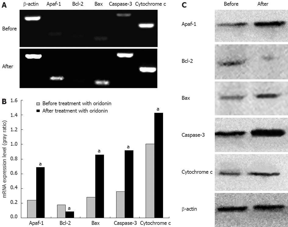
Figure 5 Changes in gene expression after treatment with 10 μg/mL oridonin for 24 h.
After treatment with oridonin for 24 h, the expression of caspase-3, cytochrome c, Apaf-1 and Bax was up-regulated, whereas that of Bcl-2 was down-regulated. A: Agarose gel electrophoresis of the reverse-transcript polymerase chain reaction products; B: Results of optical density analyses of Apaf-1/β-actin, Bcl-2/β-actin, Bax/β-actin, caspase-3/β-actin, and cytochrome c/β-actin before and after treatment with oridonin (paired t test, aP < 0.05 vs before treatment with oridonin); C: Western blotting analysis.
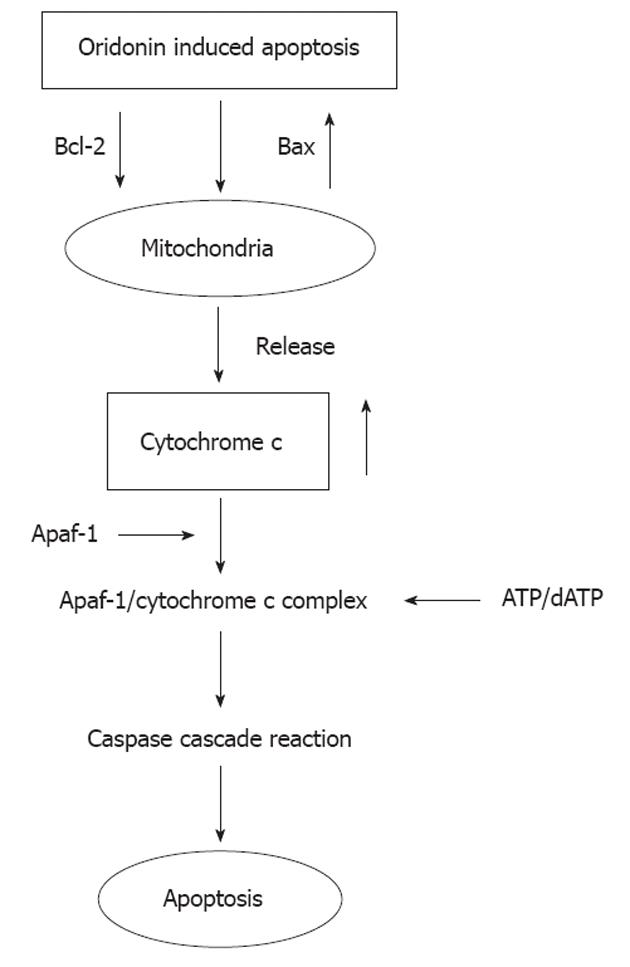
Figure 6 Possible mechanism by which oridonin induces the apoptosis of HGC-27 cells.
- Citation: Sun KW, Ma YY, Guan TP, Xia YJ, Shao CM, Chen LG, Ren YJ, Yao HB, Yang Q, He XJ. Oridonin induces apoptosis in gastric cancer through Apaf-1, cytochrome c and caspase-3 signaling pathway. World J Gastroenterol 2012; 18(48): 7166-7174
- URL: https://www.wjgnet.com/1007-9327/full/v18/i48/7166.htm
- DOI: https://dx.doi.org/10.3748/wjg.v18.i48.7166


