Copyright
©2012 Baishideng Publishing Group Co.
World J Gastroenterol. Dec 21, 2012; 18(47): 7118-7121
Published online Dec 21, 2012. doi: 10.3748/wjg.v18.i47.7118
Published online Dec 21, 2012. doi: 10.3748/wjg.v18.i47.7118
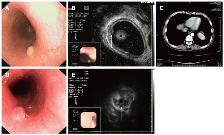
Figure 1 Clinical examination of esophageal granular cell tumors and esophageal leiomyomas.
A: Conventional gastroscopy showing a representative granular cell tumor (GCT) with white-to-yellow surface color and smooth surface; B: Endoscopic ultrasound (EUS) showing a hypoechoic lesion originating from the mucosa layer; C: Computed tomography scan showing a soft-tissue mass in the esophagus and partial narrowing of the esophageal lumen; D: Conventional gastroscopy showed that the leiomyoma had a light pink surface color with smooth surface; E: EUS showing the leiomyoma with a slightly lower echo than the esophageal GCT.
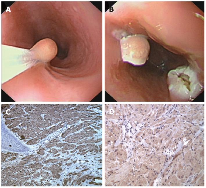
Figure 2 Clinical management and histological chrematistics of esophageal granular cell tumors.
A, B: A typical case of esophageal granular cell tumor treated by endoscopic resection; C: Histological analysis showing the lesion stained positively for S-100; D: Histological analysis showing the lesion stained positively for CD68.
- Citation: Xu GQ, Chen HT, Xu CF, Teng XD. Esophageal granular cell tumors: Report of 9 cases and a literature review. World J Gastroenterol 2012; 18(47): 7118-7121
- URL: https://www.wjgnet.com/1007-9327/full/v18/i47/7118.htm
- DOI: https://dx.doi.org/10.3748/wjg.v18.i47.7118


