Copyright
©2012 Baishideng Publishing Group Co.
World J Gastroenterol. Dec 21, 2012; 18(47): 7100-7103
Published online Dec 21, 2012. doi: 10.3748/wjg.v18.i47.7100
Published online Dec 21, 2012. doi: 10.3748/wjg.v18.i47.7100
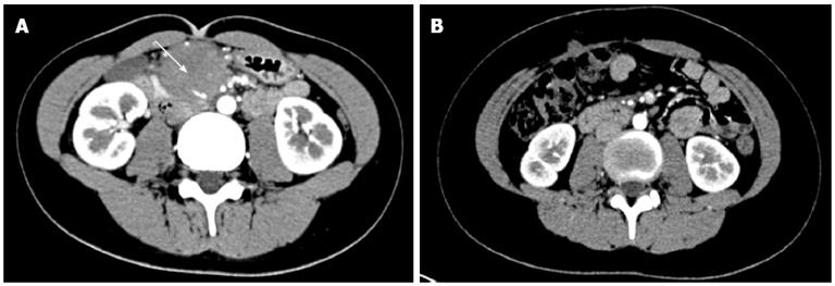
Figure 1 Photograph of abdominal computer tomography.
A: The complex soft tissue involved including the mesenteric vessels (arrow); B: Disappearance of the mass.
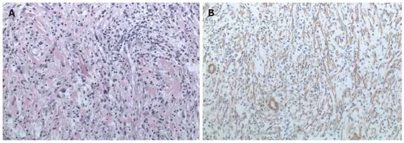
Figure 2 Photomicrograph of the tumor cells.
A: A proliferation of spindle tumor cells (hematoxylin and eosin stain, × 200); B: Positivity for smooth muscle actin (× 200).
- Citation: Tao YL, Wang ZJ, Han JG, Wei P. Inflammatory myofibroblastic tumor successfully treated with chemotherapy and nonsteroidals: A case report. World J Gastroenterol 2012; 18(47): 7100-7103
- URL: https://www.wjgnet.com/1007-9327/full/v18/i47/7100.htm
- DOI: https://dx.doi.org/10.3748/wjg.v18.i47.7100


