Copyright
©2012 Baishideng Publishing Group Co.
World J Gastroenterol. Dec 21, 2012; 18(47): 6926-6934
Published online Dec 21, 2012. doi: 10.3748/wjg.v18.i47.6926
Published online Dec 21, 2012. doi: 10.3748/wjg.v18.i47.6926
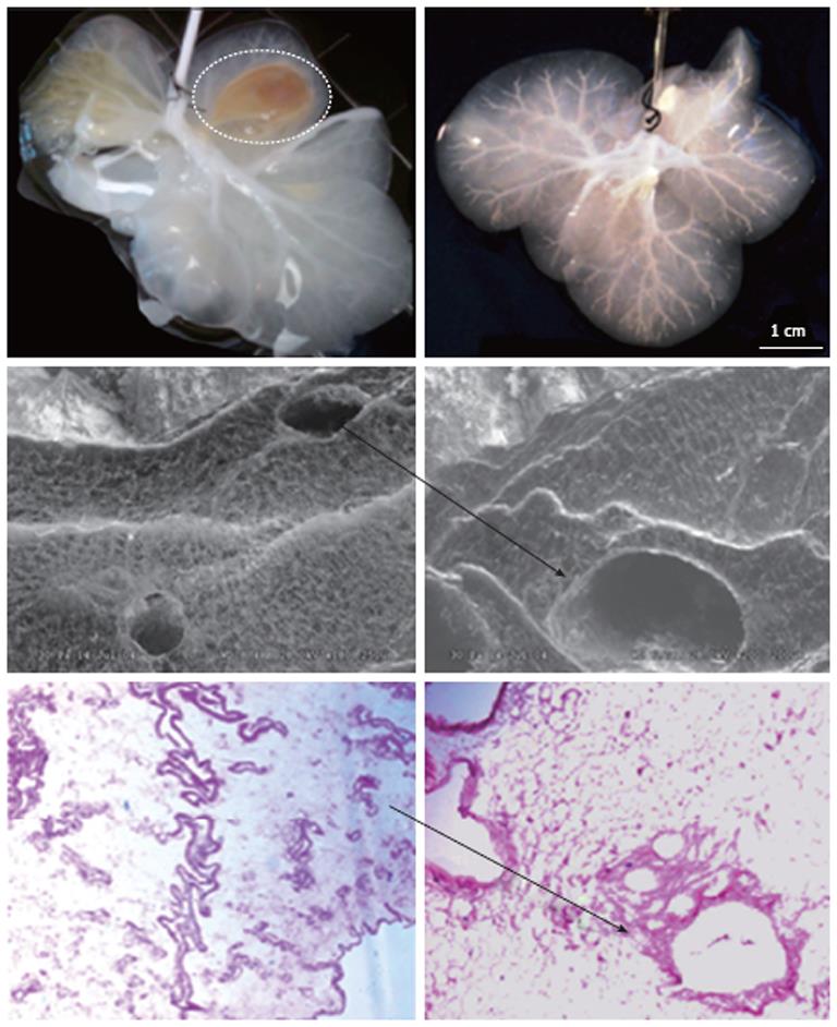
Figure 1 Gross and microscopic anatomy of acellular ferret livers.
Upper row: The liver on the left is almost entirely decellularized, however it remains a segment still cellular (interrupted line); on the left, instead, the liver is fully acellular as expression of successful decellularization; Middle row: Scanning electronic microscopic ruling out the presence of any cell remnant and showing the triad completely acellular (arrow); Lower row: Hematoxylin and eosin confirms the lack of cellular element within the remaining liver extracellular matrix (arrow).
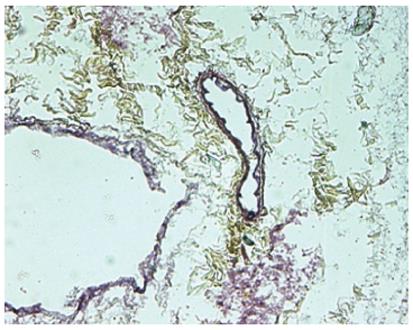
Figure 2 Movat-Pentachrome staining of acellular liver sections shows yellow staining for collagen and dark staining for elastin surrounding the vascular structures.
- Citation: Booth C, Soker T, Baptista P, Ross CL, Soker S, Farooq U, Stratta RJ, Orlando G. Liver bioengineering: Current status and future perspectives. World J Gastroenterol 2012; 18(47): 6926-6934
- URL: https://www.wjgnet.com/1007-9327/full/v18/i47/6926.htm
- DOI: https://dx.doi.org/10.3748/wjg.v18.i47.6926


