Copyright
©2012 Baishideng Publishing Group Co.
World J Gastroenterol. Nov 28, 2012; 18(44): 6379-6386
Published online Nov 28, 2012. doi: 10.3748/wjg.v18.i44.6379
Published online Nov 28, 2012. doi: 10.3748/wjg.v18.i44.6379
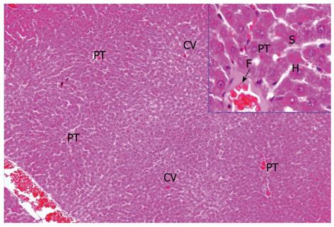
Figure 1 Liver section (hematoxylin and eosin × 100 with lateral magnification × 200) showed the normal structure of liver control.
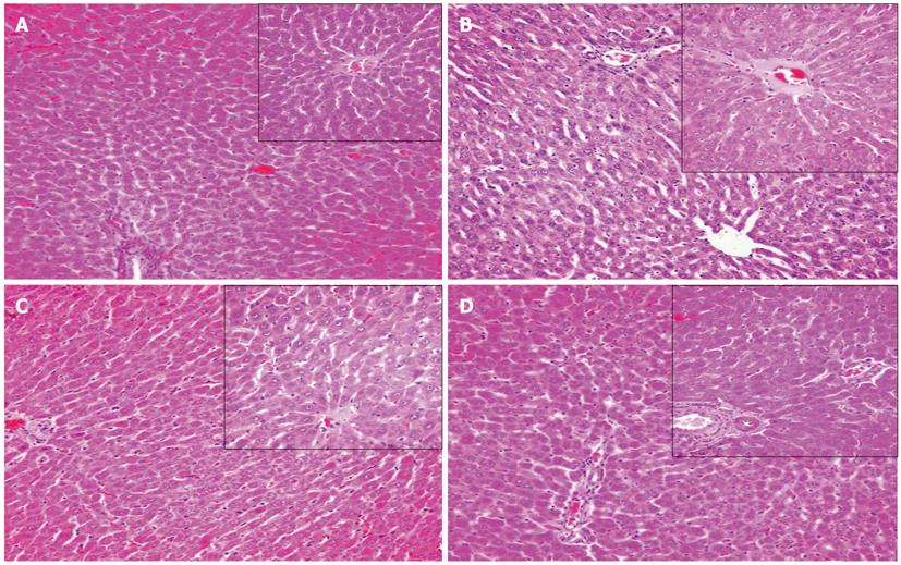
Figure 2 Liver section (hematoxylin and eosin × 200) centrilobular region and periportal region.
A: Liver section [hematoxylin and eosin (HE) × 200] centrilobular region; B: Periportal region showed severe morphological changes as a result of giving sodium nitrite; C: Liver section (HE × 200) centrilobular region; D: Periportal region of rats treated with melatonin against sodium nitrite.
Figure 3 Liver section (hematoxylin and eosin × 200) of rats.
A: Liver section [hematoxylin and eosin (HE) × 200] of rats treated with idebenone against hypox ia; B: Liver section (HE × 100 with lateral magnification × 200) of rats treated with idebenone + melatonin against sodium nitrite showing repairing of liver cells; C: Liver section (HE × 100 with lateral magnification × 200) of rats treated with arginine against sodium nitrite revealing normal hepatocyte; D: Liver section (HE × 100 with lateral magnification × 200) of rats treated with idebenone + arginine against sodium nitrite, showing hepatocytes with normal histological.
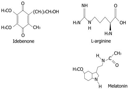
Figure 4 Chemical structures of idebenone, melatonin and arginine.
- Citation: Ali SA, Aly HF, Faddah LM, Zaidi ZF. Dietary supplementation of some antioxidants against hypoxia. World J Gastroenterol 2012; 18(44): 6379-6386
- URL: https://www.wjgnet.com/1007-9327/full/v18/i44/6379.htm
- DOI: https://dx.doi.org/10.3748/wjg.v18.i44.6379


