Copyright
©2012 Baishideng Publishing Group Co.
World J Gastroenterol. Aug 28, 2012; 18(32): 4450-4453
Published online Aug 28, 2012. doi: 10.3748/wjg.v18.i32.4450
Published online Aug 28, 2012. doi: 10.3748/wjg.v18.i32.4450
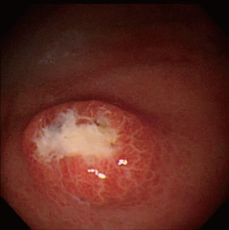
Figure 1 Endoscopic photograph showing the deep ulcer with the submucosal tumor-like margin on the greater curvature of the upper gastric body.
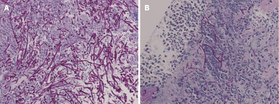
Figure 2 Biopsy demonstrated no malignancy but numerous hyphae.
A: Light micrograph of the specimen biopsied from the original ulcer demonstrating hyphae in the ulcer slough [Periodic acid-Schiff (PAS)/diastase, original magnification × 400]; B: Biopsy from the recurrent ulcer illustrating hyphae of Candida on the ulcer edge (PAS/diastase, original magnification × 400).
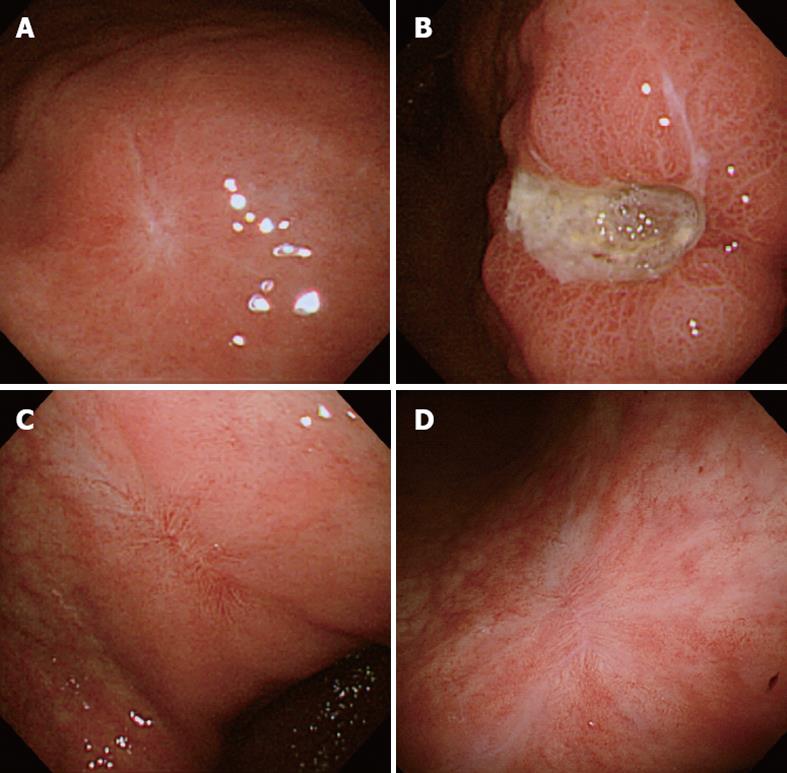
Figure 3 Endoscopic photographs.
A: The white scar of the original ulcer; B: The recurrent ulcer on the lesser curvature of the lower gastric body; C: The red scar of the recurrent ulcer; D: The transition from the red to white scar in 3 mo.
- Citation: Sasaki K. Candida-associated gastric ulcer relapsing in a different position with a different appearance. World J Gastroenterol 2012; 18(32): 4450-4453
- URL: https://www.wjgnet.com/1007-9327/full/v18/i32/4450.htm
- DOI: https://dx.doi.org/10.3748/wjg.v18.i32.4450


