Copyright
©2012 Baishideng Publishing Group Co.
World J Gastroenterol. Aug 21, 2012; 18(31): 4237-4240
Published online Aug 21, 2012. doi: 10.3748/wjg.v18.i31.4237
Published online Aug 21, 2012. doi: 10.3748/wjg.v18.i31.4237
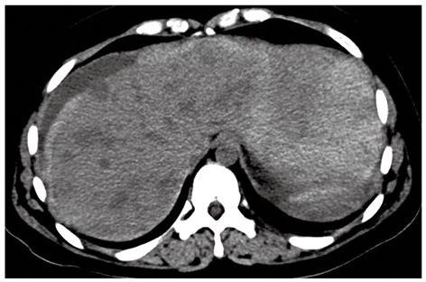
Figure 1 Axial computed tomography showing multiple hypodense nodules in the liver and hemoperitoneum around the liver and spleen.
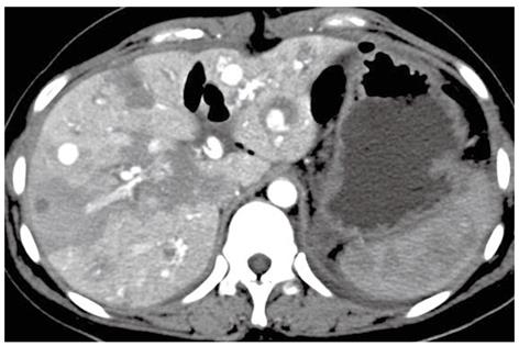
Figure 2 Contrast-enhanced computed tomography scan showing multiple hypervascular nodules similar to the enhancement of abdominal aortic aorta in the arterial phase.
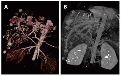
Figure 3 Volume rendering images showing multiple hypervascular nodules similar to saccular aneurysmal dilation and the presence of arterioportal shunts (white arrow).
A: Multiple hypervascular nodules similar to saccular aneurysmal dilation; B: Presence of arterioportal shunts.
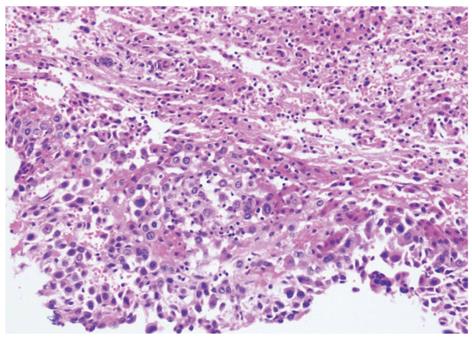
Figure 4 Histopathology showing the tumor composed of mononuclear epithelioid cells with large, irregularly shaped nuclei (hematoxylin and eosin stain × 200).
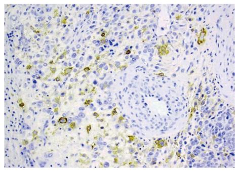
Figure 5 Immunohistochemical analysis was positive for placental alkaline phosphatase (× 200).
- Citation: Liu YH, Ma HX, Ji B, Cao DB. Spontaneous hemoperitoneum from hepatic metastatic trophoblastic tumor. World J Gastroenterol 2012; 18(31): 4237-4240
- URL: https://www.wjgnet.com/1007-9327/full/v18/i31/4237.htm
- DOI: https://dx.doi.org/10.3748/wjg.v18.i31.4237


