Copyright
©2012 Baishideng Publishing Group Co.
World J Gastroenterol. Aug 21, 2012; 18(31): 4233-4236
Published online Aug 21, 2012. doi: 10.3748/wjg.v18.i31.4233
Published online Aug 21, 2012. doi: 10.3748/wjg.v18.i31.4233
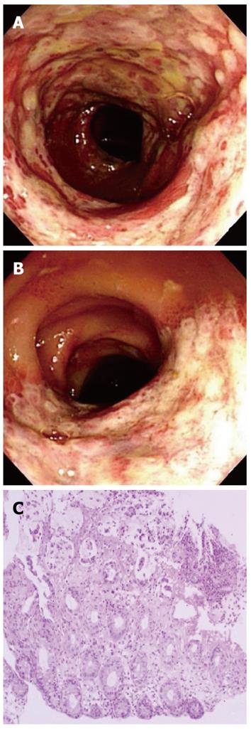
Figure 1 Colonoscopy images and pathology.
A: Mucosal hyperemic change with edema, erosion, and ulcerations and hemorrhagic friable mucosa on the sigmoid colon; B: Segmental ulceration was seen on the proximal descending colon; C: Pathological examination of the descending colon showed acute exudative colitis. Epithelial detachment was observed (hematoxylin and eosin stain, × 40).
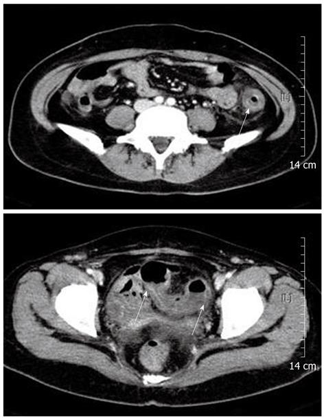
Figure 2 Abdominal computed tomography.
A: Circumferentially layered wall thickening and pericolic fat infiltration at the descending colon (arrow); B: Circumferential wall thickening and pericolic fat infiltration at the proximal sigmoid colon (arrows).
- Citation: Baik SJ, Kim TH, Yoo K, Moon IH, Cho MS. Ischemic colitis during interferon-ribavirin therapy for chronic hepatitis C: A case report. World J Gastroenterol 2012; 18(31): 4233-4236
- URL: https://www.wjgnet.com/1007-9327/full/v18/i31/4233.htm
- DOI: https://dx.doi.org/10.3748/wjg.v18.i31.4233


