Copyright
©2012 Baishideng Publishing Group Co.
World J Gastroenterol. Aug 21, 2012; 18(31): 4224-4227
Published online Aug 21, 2012. doi: 10.3748/wjg.v18.i31.4224
Published online Aug 21, 2012. doi: 10.3748/wjg.v18.i31.4224
Figure 1 Endoscopic submucosal dissection of a recurrent tumor in December 2006.
A: Pre-treatment endoscopy. A recurrent tumor 20 mm in size was detected at the site of the initial endoscopic submucosal dissection (ESD) scar by surveillance endoscopy; B: Endoscopic treatment. ESD was performed on the recurrent tumor, and an oval perforation 5 mm in length occurred at the site of extensive fibrosis during ESD; C: Endoscopic closure of the gastric perforation. The perforation was immediately observed and successfully closed with endoclips so that ESD could be continued resulting in en-bloc resection; D: Microscopic examination. A well to moderately differentiated mucosal adenocarcinoma without lymphovascular involvement was shown (hematoxylin and eosin stain, × 40); E, F: Histopathologically, an ulcer scar from the initial ESD (Ul-IIIs) was also shown [between arrowheads (E)] [hematoxylin and eosin stain, × 1 (E), × 20 (F)].
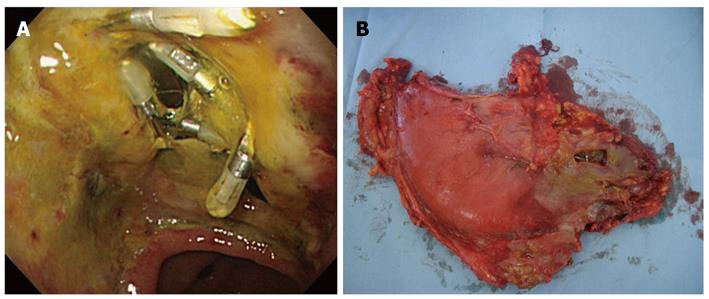
Figure 2 Dehiscence following successful endoscopic closure of the gastric perforation.
A: Endoscopy five days after endoscopic submucosal dissection (ESD). The split-open perforation previously closed successfully with endoclips during ESD was revealed; B: Surgical findings. A perforation of the gastric wall extending to the omental bursa 30 mm × 10 mm in length was seen. The perforation site was not covered with adipose tissue.
- Citation: Sekiguchi M, Suzuki H, Oda I, Yoshinaga S, Nonaka S, Saka M, Katai H, Taniguchi H, Kushima R, Saito Y. Dehiscence following successful endoscopic closure of gastric perforation during endoscopic submucosal dissection. World J Gastroenterol 2012; 18(31): 4224-4227
- URL: https://www.wjgnet.com/1007-9327/full/v18/i31/4224.htm
- DOI: https://dx.doi.org/10.3748/wjg.v18.i31.4224


