Copyright
©2012 Baishideng Publishing Group Co.
World J Gastroenterol. Jun 14, 2012; 18(22): 2877-2880
Published online Jun 14, 2012. doi: 10.3748/wjg.v18.i22.2877
Published online Jun 14, 2012. doi: 10.3748/wjg.v18.i22.2877
Figure 1 Endoscopic variceal ligation was performed to control variceal bleeding.
A: Tiny nodular varices with no bleeding were found at the duodenum second portion; B: Enhanced computed tomography scan showing large gastric varices (arrow); C: Balloon occluded retrograde transvenous obliteration was successfully performed.
Figure 2 An emergency endoscopic examination revealed a small, linear esophageal varices without evidence of recent bleeding.
A: In the second portion of the duodenum, nodular varices expanded before balloon occluded retrograde transvenous obliteration and presented with hematocystic spots; B, C: Enhanced computed tomography scan obtained 5 mo after balloon occluded retrograde transvenous obliteration revealing complete obliteration of gastric varices (black arrow); however, duodenal varices were aggravated (white arrow).
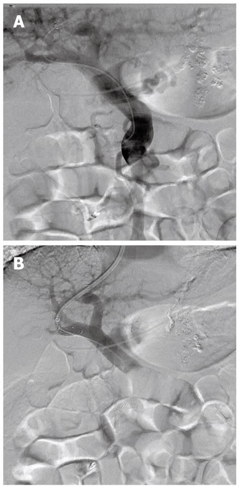
Figure 3 Transjugular intrahepatic portosystemic shunt was created according to standard procedures.
A: Portogram showing duodenal variceal flow; B: After transjugular intrahepatic portosystemic shunt and coil embolization of duodenal varices, variceal flow disappeared.
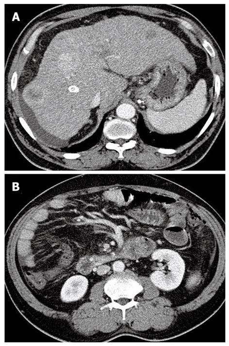
Figure 4 A follow-up computed tomography scan was obtained 21 mo after transjugular intrahepatic portosystemic shunt.
A, B: Enhanced computed tomography scan shows a still patent transjugular portosystemic shunt tract and complete improvement of duodenal varices. Note the appearance of multinodular hepatocellular carcinomas in the liver.
- Citation: Kim MJ, Jang BK, Chung WJ, Hwang JS, Kim YH. Duodenal variceal bleeding after balloon-occluded retrograde transverse obliteration: Treatment with transjugular intrahepatic portosystemic shunt. World J Gastroenterol 2012; 18(22): 2877-2880
- URL: https://www.wjgnet.com/1007-9327/full/v18/i22/2877.htm
- DOI: https://dx.doi.org/10.3748/wjg.v18.i22.2877


