Copyright
©2012 Baishideng Publishing Group Co.
World J Gastroenterol. May 21, 2012; 18(19): 2434-2437
Published online May 21, 2012. doi: 10.3748/wjg.v18.i19.2434
Published online May 21, 2012. doi: 10.3748/wjg.v18.i19.2434
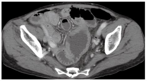
Figure 1 Computed tomography scan of the dilated and perforated small bowel.
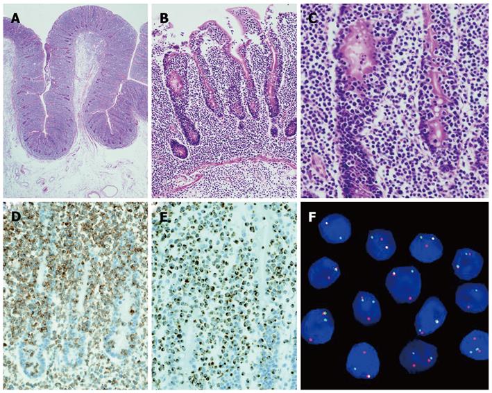
Figure 2 Pathological findings of the specimen.
A: Microscopic findings of the specimen with villous atrophy, crypt hyperplasia, and proliferation of intraepithelial lymphocytes; B: Mucosal and submucosal invasion by lymphoma cells; C: Lymphoma cells with the characteristics of intraepithelial lymphocytes; D: Expression of CD8 by intraepithelial lymphocytes; E: Expression of T cell restricted intracellular antigen-1 by intraepithelial lymphocytes; F: Examination of chromosome 8q24 (c-MYC region) using break point rearrangement probe by in situ hybridization.
- Citation: Okumura K, Ikebe M, Shimokama T, Takeshita M, Kinjo N, Sugimachi K, Higashi H. An unusual enteropathy-associated T-cell lymphoma with MYC translocation arising in a Japanese patient: A case report. World J Gastroenterol 2012; 18(19): 2434-2437
- URL: https://www.wjgnet.com/1007-9327/full/v18/i19/2434.htm
- DOI: https://dx.doi.org/10.3748/wjg.v18.i19.2434


