Copyright
©2012 Baishideng Publishing Group Co.
World J Gastroenterol. Mar 14, 2012; 18(10): 1059-1066
Published online Mar 14, 2012. doi: 10.3748/wjg.v18.i10.1059
Published online Mar 14, 2012. doi: 10.3748/wjg.v18.i10.1059
Figure 1 Effects of rebamipide on diclofenac-induced small intestinal permeability in mice.
Values are mean ± SEM of data obtained from 8 mice in each group. bP < 0.01 compared with the diclofenac group.
Figure 2 Transmission electron microscopic appearances of diclofenac-induced small intestinal injuries in mice (original magnification × 20 000).
A: Control group; B: Diclofenac group; C: Rebamipide group (400 mg/kg). In the diclofenac group, partial deformation of intestinal epithelial cells, intestinal microvillus reduction, disarrangement of the epithelial surface and broader junctional complexes, tight junction opening were seen. Rebamipide group showed regular and intensive microvillus, and ameliorated tight junction when compared with the diclofenac group.
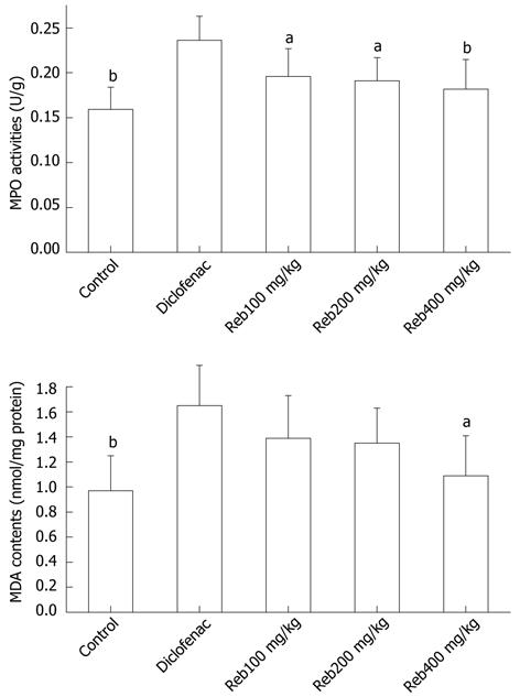
Figure 3 Effects of rebamipide on small intestinal malondialdehyde content and myeloperoxidase activity.
Values are mean ± SEM of data obtained from 8 mice in each group. aP < 0.05, bP < 0.01 compared with the diclofenac group. MDA: Malondialdehyde; MPO: Myeloperoxidase.
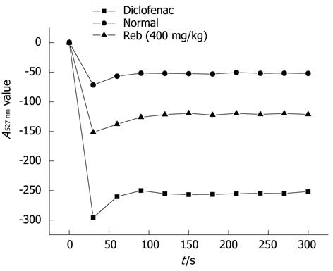
Figure 4 Effects of rebamipide on diclofenac-induced liver mitochondrial membrane potential.
In the control group, the fluorescence intensity was reduced within 30 s, and then began to rise and reached a steady state within 90 s. The fluorescence intensity reduction in the diclofenac group was smaller than that in the control group, indicating that the liver mitochondrial membrane potential was decreased by diclofenac administration. The reduction in the fluorescence intensity in the rebamipide group was greater than that in the diclofenac group indicating that rebamipide improved the impairment in mitochondrial function induced by diclofenac.
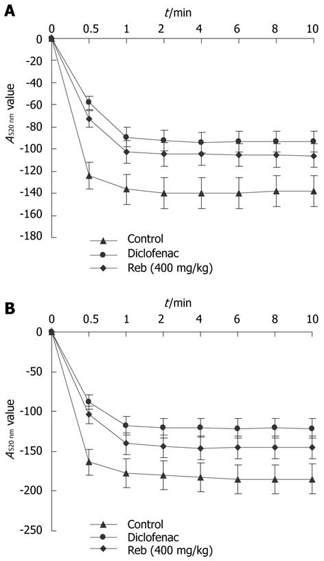
Figure 5 Effects of rebamipide on diclofenac-induced liver mitochondrial swelling in mice.
A: After adding the reaction buffer, the absorbance at 520 nm in the control mitochondria declined rapidly. The decrease was smaller in the presence of diclofenac compared with that in the control, demonstrating that liver mitochondrial dysfunction was induced by diclofenac administration. This reduction in absorbance was significantly increased in the presence of rebamipide, indicating that rebamipide improved impaired mitochondrial function; B: After adding 0.3 mmol/L CaC12 reaction buffer, the absorbance at 520 nm in the control mitochondria declined rapidly, suggesting significant swelling of mitochondria. The decrease was smaller in the presence of diclofenac compared with that in the control, demonstrating that liver mitochondrial dysfunction was induced by diclofenac administration. This reduction was significantly increased in the presence of rebamipide, indicating that rebamipide improved impaired mitochondrial function.
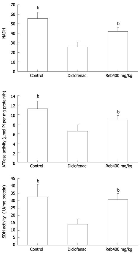
Figure 6 Effects of rebamipide on diclofenac-induced decreases in liver mitochondrial nicotinamide adenine dinucleotide-reduced levels, succinate dehydrogenase and ATPase activities in mice.
Values are mean ± SEM of data obtained from 8 mice in each group. bP < 0.01 compared with the diclofenac group. SDH: Succinate dehydrogenase; NADH: Nicotinamide adenine dinucleotide-reduced.
-
Citation: Diao L, Mei Q, Xu JM, Liu XC, Hu J, Jin J, Yao Q, Chen ML. Rebamipide suppresses diclofenac-induced intestinal permeability
via mitochondrial protection in mice. World J Gastroenterol 2012; 18(10): 1059-1066 - URL: https://www.wjgnet.com/1007-9327/full/v18/i10/1059.htm
- DOI: https://dx.doi.org/10.3748/wjg.v18.i10.1059


