Copyright
©2011 Baishideng Publishing Group Co.
World J Gastroenterol. Sep 7, 2011; 17(33): 3836-3841
Published online Sep 7, 2011. doi: 10.3748/wjg.v17.i33.3836
Published online Sep 7, 2011. doi: 10.3748/wjg.v17.i33.3836
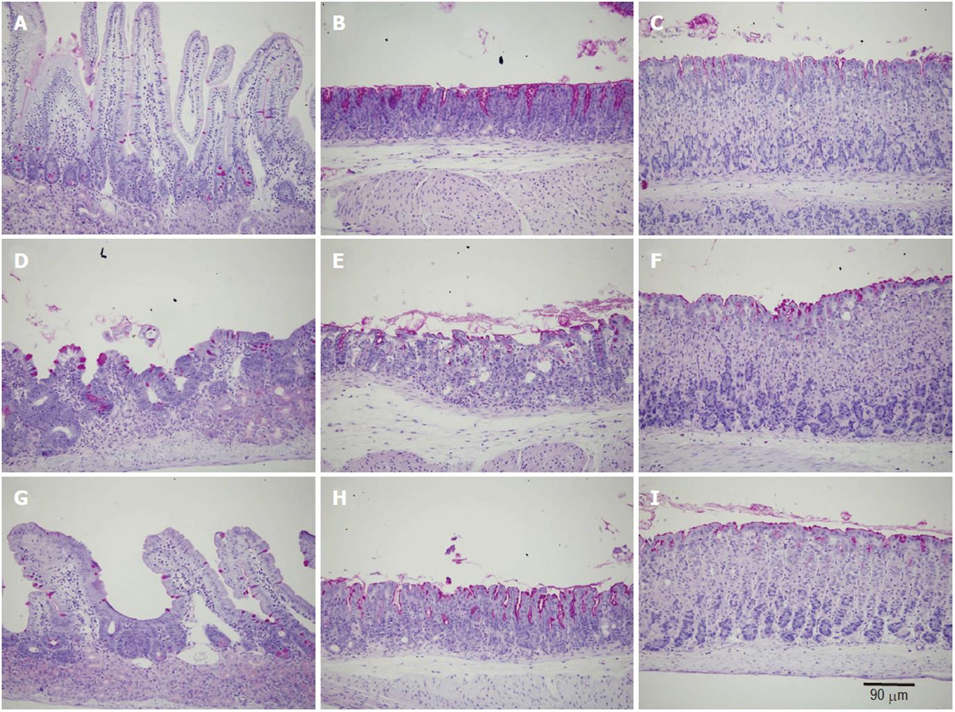
Figure 1 Light micrographs showing tissue sections obtained from the duodenum (A, D, G), pyloric antrum (B, E, H) and gastric corpus (C, F, I) of control (A-C), doxorubicin-treated (D-F) and ghrelin-plus-doxorubicin-treated (G-I) mice.
All tissue sections were stained with periodic acid schiff and hematoxylin. Note that while there are apparent mucosal changes in D and E due to doxorubicin treatment, the tissues in G and H are more or less similar to control.
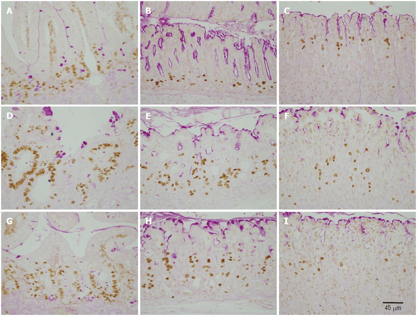
Figure 2 Immunohistochemical analysis of S-phase cells using anti-5’-bromo-2’-deoxyuridine antibody and tissue sections obtained from the duodenum (A, D, G), pyloric antrum (B, E, H) and gastric corpus (C, F, I) of control (A-C), doxorubicin-treated (D-F) and ghrelin/doxorubicin-treated (G-I) mice.
Note that BrdU-labeled cells (brown nuclei) of doxorubicin-treated tissues in D, E and F appear more expanded when compared with control tissues in A, B and C. BrdU-labeled cells in G and and H are still more expanded than in the control, but less prominent than in D and E.
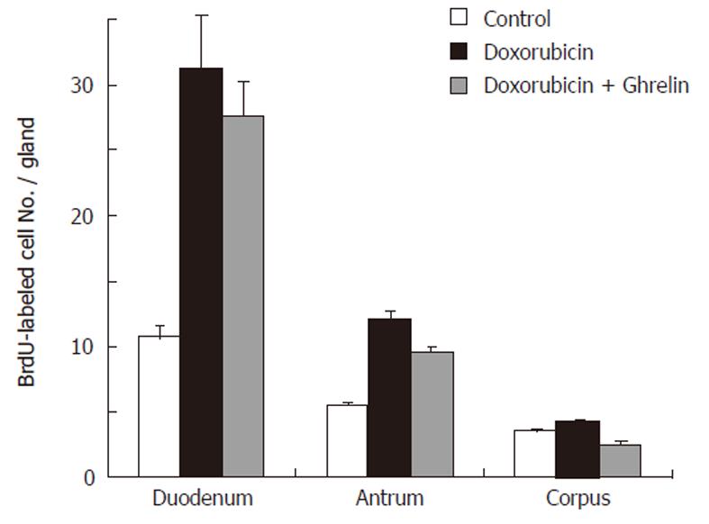
Figure 3 Analysis of 5’-bromo-2’-deoxyuridine-labeled cell counts in the duodenum, antrum and corpus of control, doxorubicin-treated and ghrelin/doxorubicin-treated mice.
- Citation: Fahim MA, Kataya H, El-Kharrag R, Amer DA, al-Ramadi B, Karam SM. Ghrelin attenuates gastrointestinal epithelial damage induced by doxorubicin. World J Gastroenterol 2011; 17(33): 3836-3841
- URL: https://www.wjgnet.com/1007-9327/full/v17/i33/3836.htm
- DOI: https://dx.doi.org/10.3748/wjg.v17.i33.3836


