Copyright
©2011 Baishideng Publishing Group Co.
World J Gastroenterol. Jul 21, 2011; 17(27): 3263-3266
Published online Jul 21, 2011. doi: 10.3748/wjg.v17.i27.3263
Published online Jul 21, 2011. doi: 10.3748/wjg.v17.i27.3263
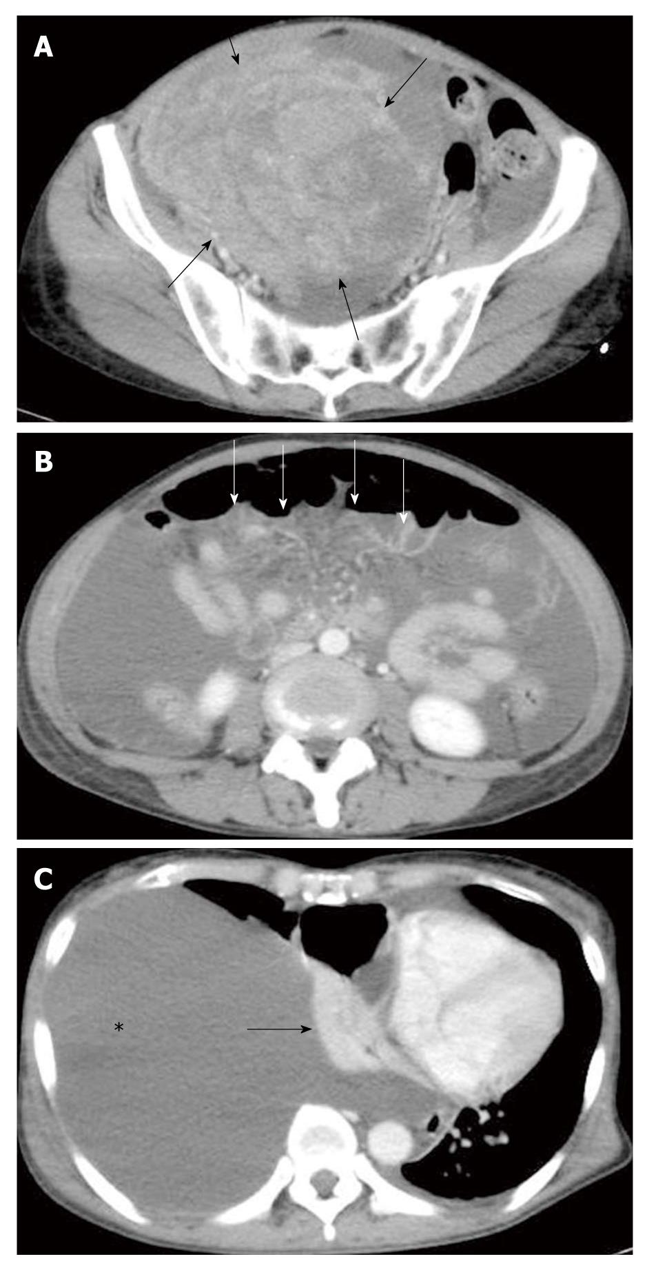
Figure 1 Computed tomography.
A: A huge round mass in the pelvis suggesting a bilateral ovarian tumor (black arrows); B: Massive ascites and thickness of omentum (white allows) suggesting peritoneal dissemination; C: Right pleural effusion with atelectasis (black allow) and left-side compression of the mediastinum.
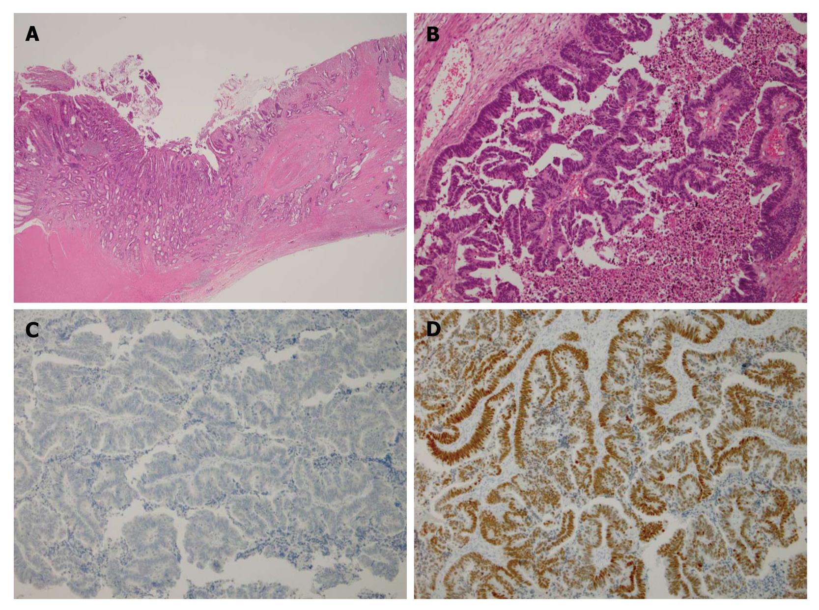
Figure 2 Microscopic features.
A: Well-differentiated adenocarcinoma compatible with primary colon cancer; hematoxylin and eosin staining; B: The ovarian tumor is composed of tumor cells compatible with metastasis of colon cancer; C: Cytokeratin 7 showing negative result of staining in the ovarian tumor; D: CDX-2 showing staining in tumor cells in the ovary.
- Citation: Maeda H, Okabayashi T, Hanazaki K, Kobayashi M. Clinical experience of Pseudo-Meigs’ Syndrome due to colon cancer. World J Gastroenterol 2011; 17(27): 3263-3266
- URL: https://www.wjgnet.com/1007-9327/full/v17/i27/3263.htm
- DOI: https://dx.doi.org/10.3748/wjg.v17.i27.3263


