Copyright
©2011 Baishideng Publishing Group Co.
World J Gastroenterol. Jul 21, 2011; 17(27): 3220-3228
Published online Jul 21, 2011. doi: 10.3748/wjg.v17.i27.3220
Published online Jul 21, 2011. doi: 10.3748/wjg.v17.i27.3220
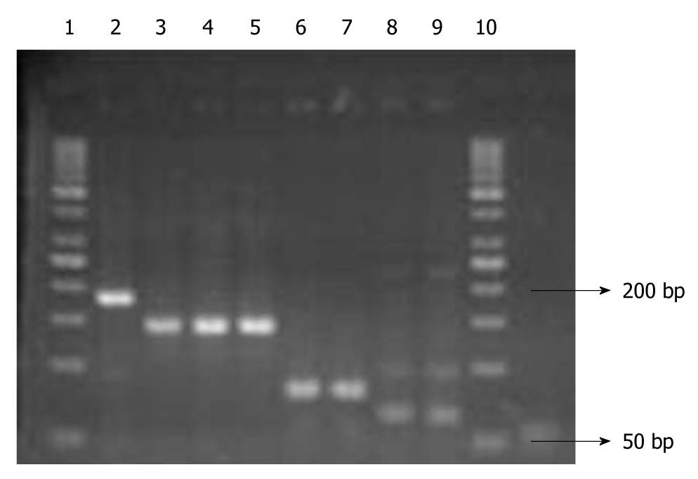
Figure 1 Polymerase chain reaction products of genes.
1: Ladder (50 bp); 2: Lymphatic vessel endothelial hyaluronan receptor-1 (184 bp), gastric tissue; 3-5: GAPDH (143 bp), in colon, stomach and pancreas, respectively; 6, 7: CD44 (80 bp) in stomach and colon; 8, 9: Vascular endothelial growth factor receptor-3 (63 bp) in stomach and colon; 10: ladder (50 bp).
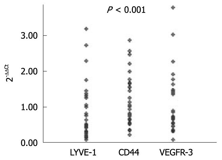
Figure 2 Lymphatic vessel endothelial hyaluronan receptor-1, CD44 and vascular endothelial growth factor receptor-3 expression levels are increased in tumor tissues as compared to normal tissues in patients with gastric cancer (P < 0.
001). LYVE-1: Lymphatic vessel endothelial hyaluronan receptor-1; VEGFR-3: Vascular endothelial growth factor receptor-3
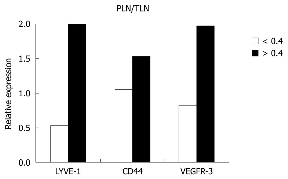
Figure 3 The positive lymph nodes/total lymph nodes ratios and the relative expression levels.
The expression levels of lymphatic vessel endothelial hyaluronan receptor-1 (LYVE-1) are increased in conjunction with increased positive lymph nodes (PLN)/total lymph nodes (TLN) ratio (P = 0.003). VEGFR-3: Vascular endothelial growth factor receptor-3.
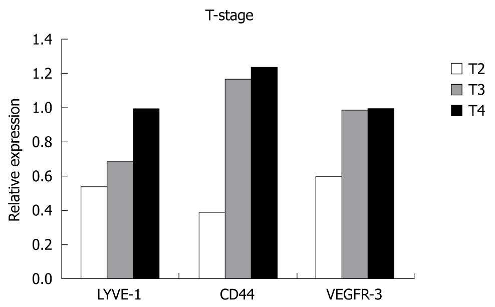
Figure 4 The relative expression levels and T-stage.
No correlation was found between expression levels and T-stage. LYVE-1: Lymphatic vessel endothelial hyaluronan receptor-1; VEGFR-3: Vascular endothelial growth factor receptor-3.
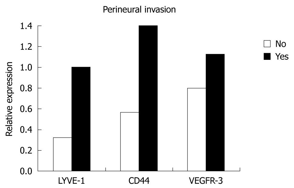
Figure 5 The relative expression levels and perineural invasion.
The expression levels of lymphatic vessel endothelial hyaluronan receptor-1 (LYVE-1) and CD44 were significantly increased with the presence of perineural invasion. VEGFR-3: Vascular endothelial growth factor receptor-3.
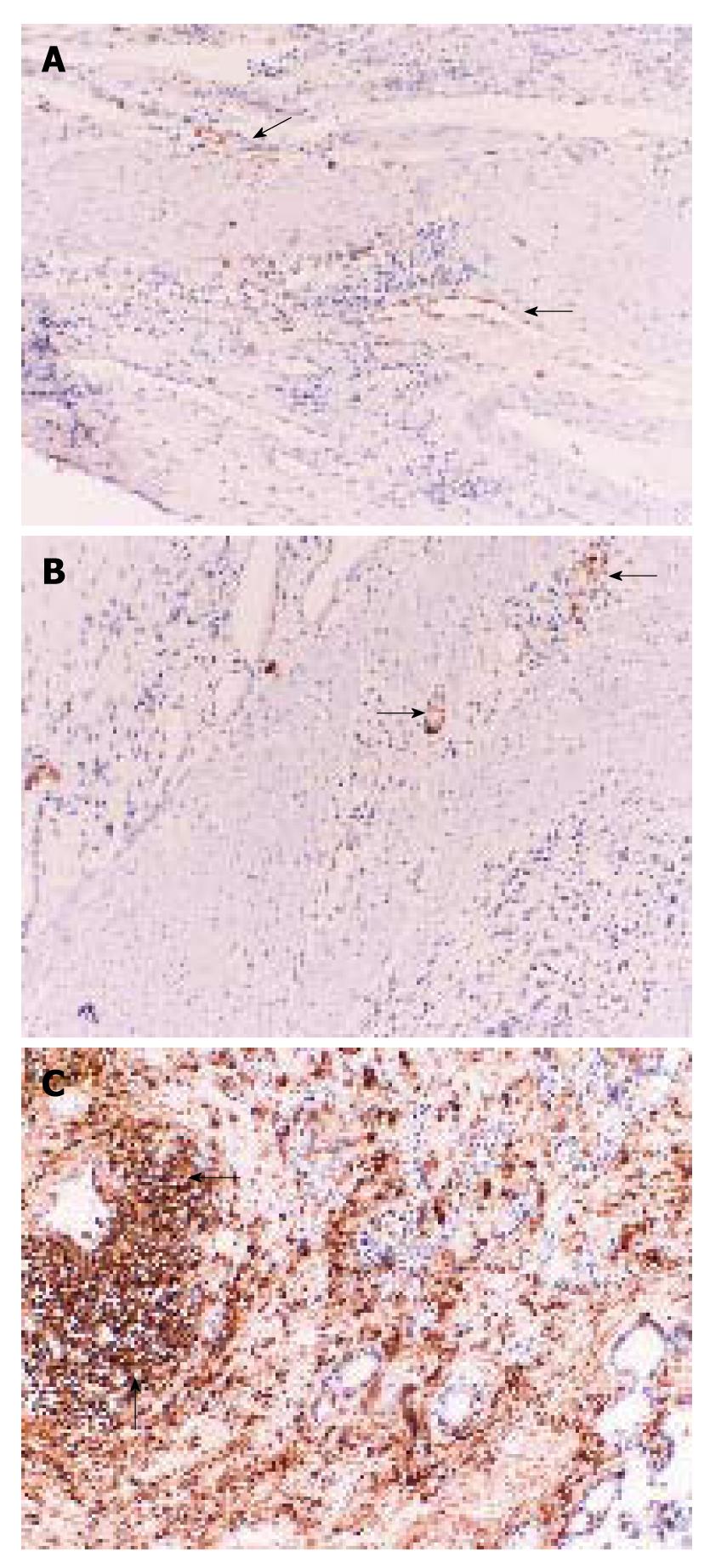
Figure 6 Expressions of lymphatic vessel endothelial hyaluronan receptor-1, vascular endothelial growth factor receptor-3 and CD44 by immunohistochemistry.
A: Arrows indicate the lymphatic vessel endothelial hyaluronan receptor-1-positive lymphatic vessels in gastric tumor (× 100); B: Arrows indicate vascular endothelial growth factor receptor-3-3-positive lymphatic vessels in gastric tumor (× 100); C: Arrows indicate CD44-positive lymphoid cells in gastric tumor (× 100).
- Citation: Ozmen F, Ozmen MM, Ozdemir E, Moran M, Seçkin S, Guc D, Karaagaoglu E, Kansu E. Relationship between LYVE-1, VEGFR-3 and CD44 gene expressions and lymphatic metastasis in gastric cancer. World J Gastroenterol 2011; 17(27): 3220-3228
- URL: https://www.wjgnet.com/1007-9327/full/v17/i27/3220.htm
- DOI: https://dx.doi.org/10.3748/wjg.v17.i27.3220


