Copyright
©2011 Baishideng Publishing Group Co.
World J Gastroenterol. Jun 7, 2011; 17(21): 2618-2622
Published online Jun 7, 2011. doi: 10.3748/wjg.v17.i21.2618
Published online Jun 7, 2011. doi: 10.3748/wjg.v17.i21.2618
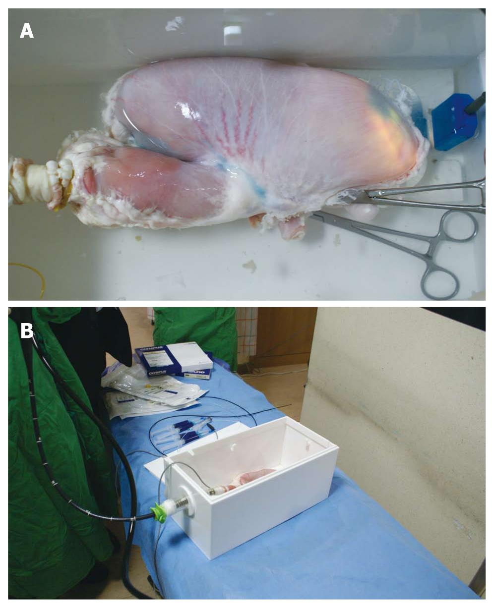
Figure 1 Porcine stomach model for endoscopic submucosal dissection.
The stomach was immersed in water of the frame for detection of air leakage. The duodenum was sealed with Kelly. The electrical plate was attached to the bottom of the stomach. Bluish discoloration with light illumination was seen at the stomach antrum area (A); the endoscope was introduced through the overtube, which was fixed to the frame developed for endoscopic submucosal dissection tutoring (B).
- Citation: Kim EY, Jeon SW, Kim GH. Chicken soup for teaching and learning ESD. World J Gastroenterol 2011; 17(21): 2618-2622
- URL: https://www.wjgnet.com/1007-9327/full/v17/i21/2618.htm
- DOI: https://dx.doi.org/10.3748/wjg.v17.i21.2618


