Copyright
©2011 Baishideng Publishing Group Co.
World J Gastroenterol. Apr 14, 2011; 17(14): 1817-1824
Published online Apr 14, 2011. doi: 10.3748/wjg.v17.i14.1817
Published online Apr 14, 2011. doi: 10.3748/wjg.v17.i14.1817
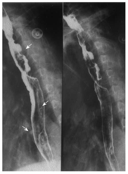
Figure 1 Sixty four-year-old male with three esophageal lesions.
The first was a medullary lesion (↑) located at the upper, the other two were small ulcerative lesions (↑) located at the middle-lower segment, and all were squamous cell carcinoma, pathologically proved.
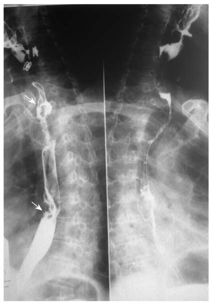
Figure 2 Fifty eight-year-old male with two esophageal lesions.
The first was a medullary lesion located at the upper segment, with stenosis and rough rigid margins (↑); the other was a proliferative nodule located at the middle segment, with disruptive mucosa (↑).
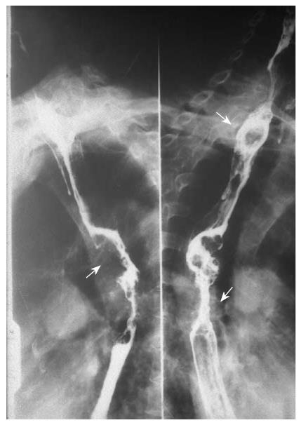
Figure 3 Sixty three-year-old male with two esophageal lesions.
The first was a proliferative nodule located at the upper (↑), the other was a medullary lesion located at the middle segment (↑), and all were squamous cell carcinoma, pathologically proved.
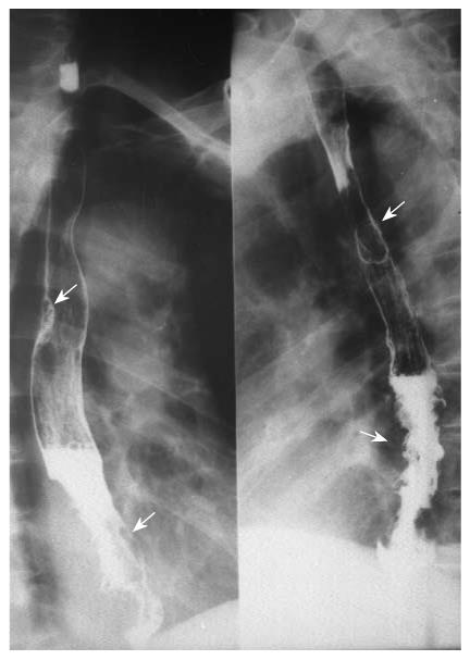
Figure 4 Fifty six-year-old female with two esophageal lesions.
The first was a small proliferative nodule located at the middle (↑), the other was a medullary lesion located at the lower segment (↑), and all were squamous cell carcinoma, pathologically proved.
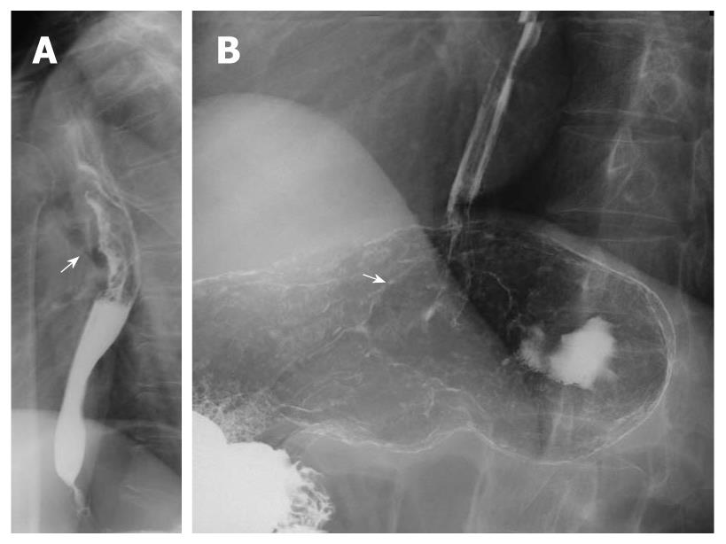
Figure 5 Forty nine-year-old male with esophageal-gastric multiple primary carcinoma.
The esophageal lesion was ulcerative, located at the middle segment (↑); the gastric lesion was proliferative, located at the cardia (↑), with distal esophageal infiltration.
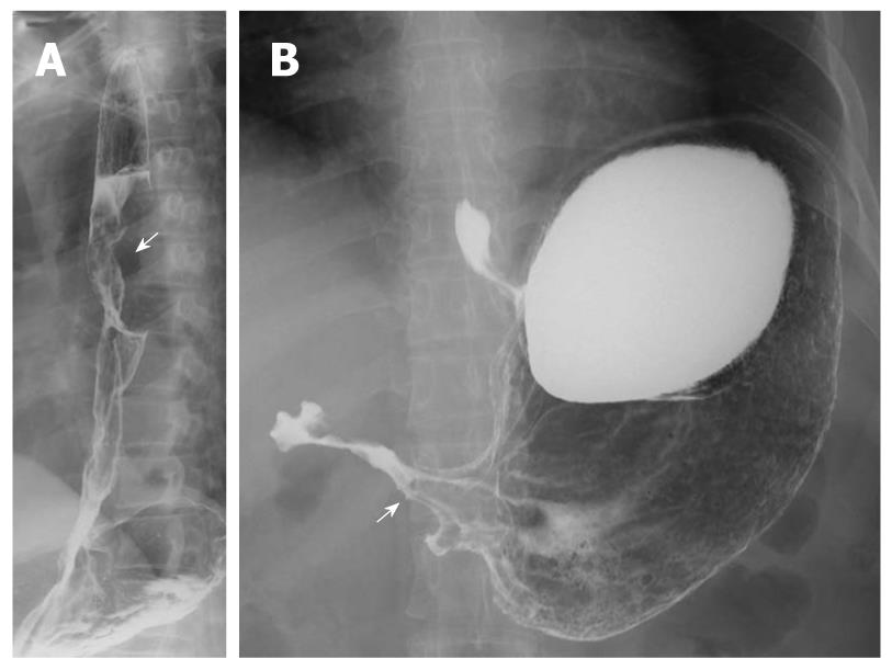
Figure 6 Fifty seven-year-old male with esophageal-gastric multiple primary carcinoma.
The esophageal lesion was proliferative located at the middle segment (↑); the gastric lesion was infiltrative located at the antrum (↑).
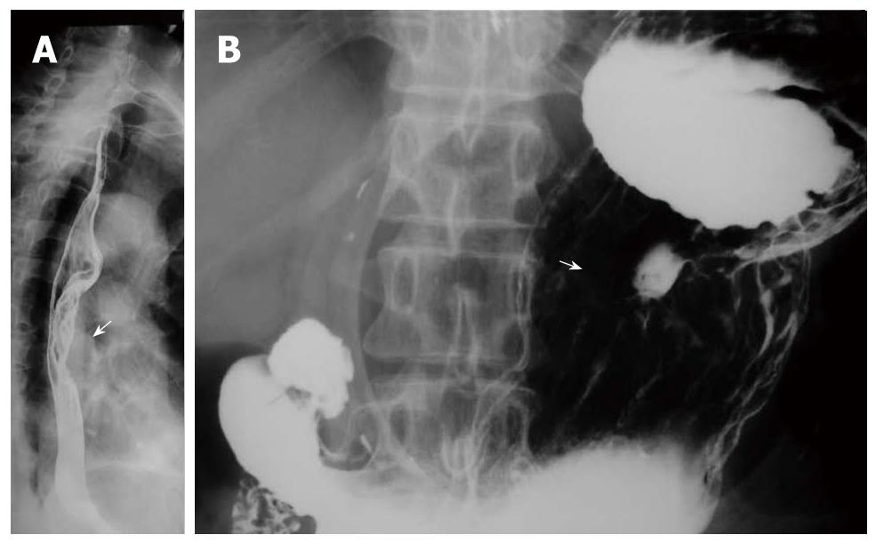
Figure 7 Forty six-year-old female with esophageal-gastric multiple primary carcinoma.
The esophageal lesion was proliferative located at the middle (↑); the gastric lesion was ulcerative located at the body (↑).
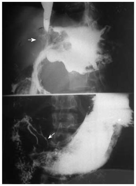
Figure 8 Fifty nine-year-old male with two gastric lesions.
The main lesion was proliferative (↑) located at the cardia, with fundus infiltration; the additional one was also a proliferative lesion located at the antrum, with disruptive mucosa (↑). All were adenocarcinoma, pathologically proved.
- Citation: Yang ZH, Gao JB, Yue SW, Guo H, Yang XH. X-ray diagnosis of synchronous multiple primary carcinoma in the upper gastrointestinal tract. World J Gastroenterol 2011; 17(14): 1817-1824
- URL: https://www.wjgnet.com/1007-9327/full/v17/i14/1817.htm
- DOI: https://dx.doi.org/10.3748/wjg.v17.i14.1817


