Copyright
©2011 Baishideng Publishing Group Co.
World J Gastroenterol. Mar 14, 2011; 17(10): 1358-1361
Published online Mar 14, 2011. doi: 10.3748/wjg.v17.i10.1358
Published online Mar 14, 2011. doi: 10.3748/wjg.v17.i10.1358
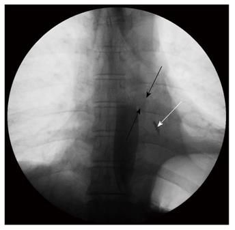
Figure 1 Barium esophagography showing the esophageal orifice of fistula (black arrows) and some barium in the left lower lobe (white arrow) demonstrating the downward direction of the fistula from esophagus to the left lower lobe bronchus.
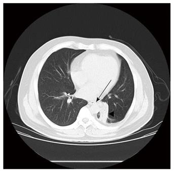
Figure 2 Preoperative chest computed tomography scan demonstrating abnormal communication between the lower third of esophagus and the left segmental bronchus (arrow) and a mass measuring 3.
0 cm in diameter with irregular borders encircling the basal segmental bronchi of the left lower lobe caused by bronchoesophageal fistula.
- Citation: Zhang BS, Zhou NK, Yu CH. Congenital bronchoesophageal fistula in adults. World J Gastroenterol 2011; 17(10): 1358-1361
- URL: https://www.wjgnet.com/1007-9327/full/v17/i10/1358.htm
- DOI: https://dx.doi.org/10.3748/wjg.v17.i10.1358


