Copyright
©2011 Baishideng Publishing Group Co.
World J Gastroenterol. Mar 14, 2011; 17(10): 1343-1353
Published online Mar 14, 2011. doi: 10.3748/wjg.v17.i10.1343
Published online Mar 14, 2011. doi: 10.3748/wjg.v17.i10.1343
Figure 1 Pancreas-preserving total duodenectomy technique.
A: The duodenum is separated from the head of the pancreas by cutting the branches of the pancreaticoduodenal arcade vessels. The choledochal trunk is saved and only the membrane of the major papilla is shaved sharply. The accessory pancreatic duct is ligated and cut; B: Papillotomy is performed on the major papilla at 0, and then a catheter is inserted into the main pancreatic duct for stenting; C: Bilio-jejunal reconstruction. The edge of the common bile duct is sewn to a small opening on the jejunum with 4-0 absorbable knotted sutures; D:The final reconstruction schema of the alimentary tract.
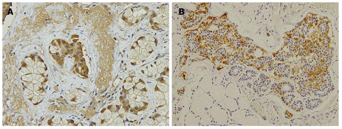
Figure 2 Hyperplasia (A) and a cluster of gastrin-producing cells (B) in the duodenal Brunner’s glands (in patient No.
12, who underwent pancreas-preserving total duodenectomy for numerous duodenal microgastrinomas) were detected by immunohistochemical gastrin staining.
Figure 3 Survival curve of 16 patients with multiple endocrine neoplasia type 1 and neuroendocrine tumors.
A: Overall survival after initial resection surgery. The survival rate at 10 years was 90.9%; B:Gastrinoma-free survival after initial resection surgery. The survival rate at 10 years was 82.0%.
- Citation: Imamura M, Komoto I, Ota S, Hiratsuka T, Kosugi S, Doi R, Awane M, Inoue N. Biochemically curative surgery for gastrinoma in multiple endocrine neoplasia type 1 patients. World J Gastroenterol 2011; 17(10): 1343-1353
- URL: https://www.wjgnet.com/1007-9327/full/v17/i10/1343.htm
- DOI: https://dx.doi.org/10.3748/wjg.v17.i10.1343


