Copyright
©2011 Baishideng Publishing Group Co.
World J Gastroenterol. Mar 14, 2011; 17(10): 1276-1285
Published online Mar 14, 2011. doi: 10.3748/wjg.v17.i10.1276
Published online Mar 14, 2011. doi: 10.3748/wjg.v17.i10.1276
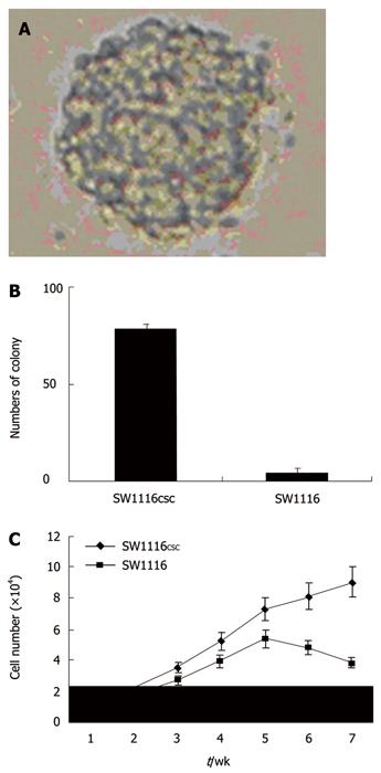
Figure 1 Spheres (A), sphere formation assay (B), and proliferation assay (C) of SW1116 cells.
Figure 2 Flow cytometry analysis showing the expressions of CD133 and CD29 (A) and the positive rates of CD133 and CD29 (B) in SW1116 cells.
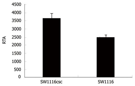
Figure 3 Telomerase activities in SW1116csc and SW1116 cells.
RTA: Relative telomerase activities.
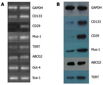
Figure 4 Expressions of CD133, CD29, Musashi-1, TERT, ABCG2, Oct-4 and Sca-1 genes (A) and CD133, CD29, Musashi-1, ABCG2 and TERT proteins (B) in SW1116csc and SW1116 cells (left: SW1116 cells, right: SW1116csc).
GAPDH: Glyceraldehyde-3-phosphate dehydrogenase; Mus-1: Musashi-1.
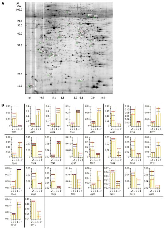
Figure 5 Two-dimensional gel electrophoresis profile (A) and histogram (B) showing expression levels of 26 protein spots in SW1116 cells and SW1116csc.
- Citation: Zou J, Yu XF, Bao ZJ, Dong J. Proteome of human colon cancer stem cells: A comparative analysis. World J Gastroenterol 2011; 17(10): 1276-1285
- URL: https://www.wjgnet.com/1007-9327/full/v17/i10/1276.htm
- DOI: https://dx.doi.org/10.3748/wjg.v17.i10.1276


