Copyright
©2010 Baishideng.
World J Gastroenterol. Jan 28, 2010; 16(4): 526-530
Published online Jan 28, 2010. doi: 10.3748/wjg.v16.i4.526
Published online Jan 28, 2010. doi: 10.3748/wjg.v16.i4.526
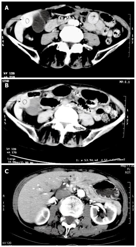
Figure 1 Contrast CT scan of the abdomen.
A, B: CT scan of the abdomen showing a 2.5-cm tumor in segment 5 of the liver (arrow); C: CT scan of the abdomen showing a 3-cm tumor in the left lateral segment of the liver (arrow).
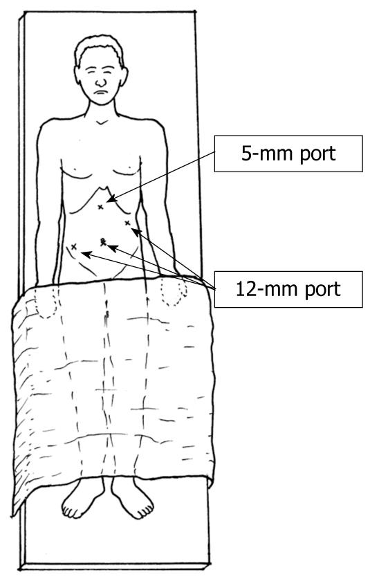
Figure 2 Port placement for laparoscopic hepatectomy.
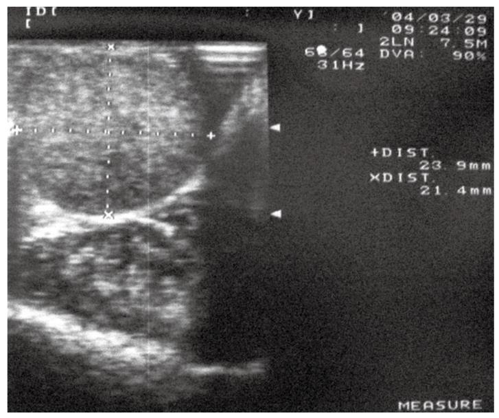
Figure 3 Intraoperative ultrasound scan of the liver showing a tumor measuring 23.
9 mm × 21.4 mm in segment 5.
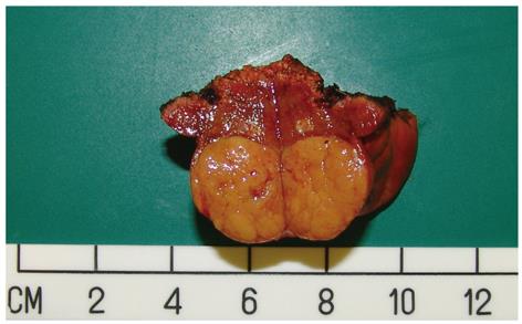
Figure 4 The 2.
5-cm resected tumor that originated in segment 5 of the liver after laparoscopic wedge excision.
- Citation: Cheung TT, Ng KKC, Poon RTP, Chan SC, Lo CM, Fan ST. A case of laparoscopic hepatectomy for recurrent hepatocellular carcinoma. World J Gastroenterol 2010; 16(4): 526-530
- URL: https://www.wjgnet.com/1007-9327/full/v16/i4/526.htm
- DOI: https://dx.doi.org/10.3748/wjg.v16.i4.526


