Copyright
©2010 Baishideng.
World J Gastroenterol. Jan 28, 2010; 16(4): 522-525
Published online Jan 28, 2010. doi: 10.3748/wjg.v16.i4.522
Published online Jan 28, 2010. doi: 10.3748/wjg.v16.i4.522
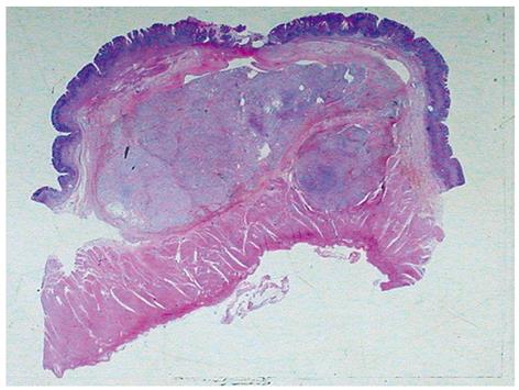
Figure 1 Low power view showing the tumor predominantly located in the submucosa with focal ulceration of the adjacent mucosa.
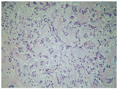
Figure 2 Microscopy showing the tumor cells mostly arranged as small nests commonly related to variably sized vessels, with abundant extracellular material and mucinous or collagenous characteristics.
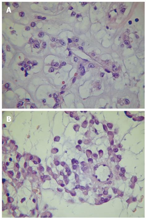
Figure 3 Neoplastic cells showing eosinophilic and clear epithelioid characteristics and perivascular arrangement (A) and focal rhabdoid features (B).
- Citation: Mitteldorf CATDS, Birolini D, Camara-Lopes LHD. A perivascular epithelioid cell tumor of the stomach: An unsuspected diagnosis. World J Gastroenterol 2010; 16(4): 522-525
- URL: https://www.wjgnet.com/1007-9327/full/v16/i4/522.htm
- DOI: https://dx.doi.org/10.3748/wjg.v16.i4.522


