Copyright
©2010 Baishideng.
World J Gastroenterol. Jan 28, 2010; 16(4): 439-444
Published online Jan 28, 2010. doi: 10.3748/wjg.v16.i4.439
Published online Jan 28, 2010. doi: 10.3748/wjg.v16.i4.439
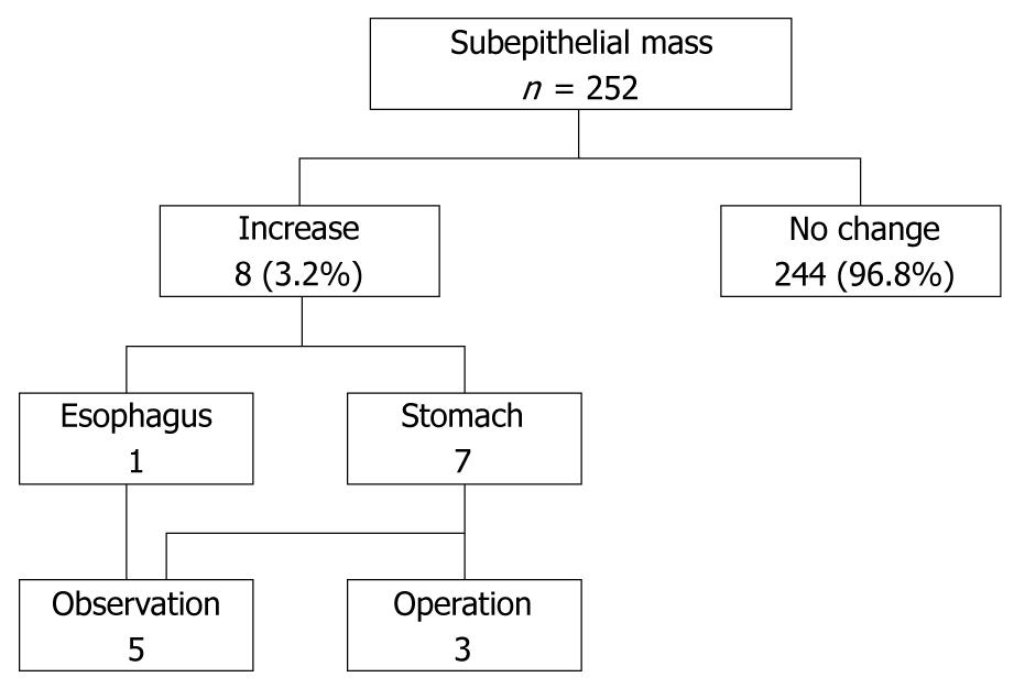
Figure 1 Clinical course of subepithelial masses.
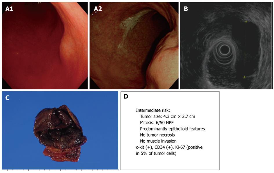
Figure 2 Endoscopic, endoscopic ultrasonography (EUS), and gross findings of gastrointestinal stromal tumors (GISTs).
A: Endoscopic view of a round subepithelial mass with a significant interval change; B: EUS shows an ovoid, homogeneous, hypoechoic mass in the fourth gastric wall layer; C: Gross findings of wedge resection reveal a soft, well-defined mass measuring 4.3 cm × 2.7 cm; D: Malignant potential.
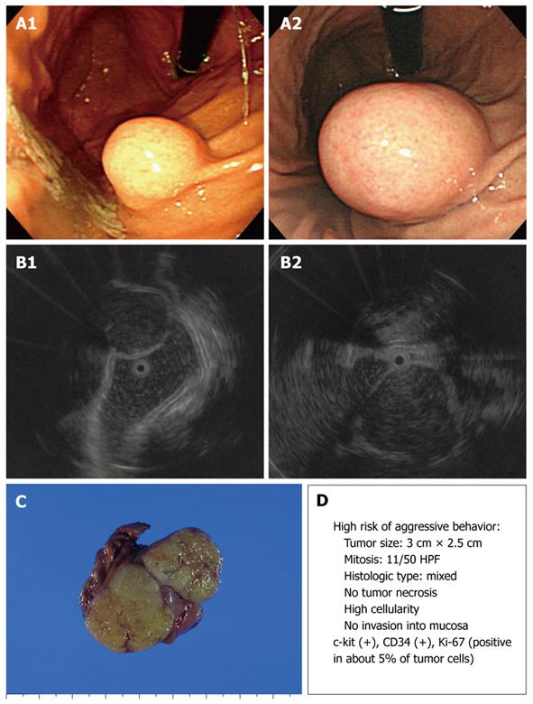
Figure 3 Endoscopic, EUS, and gross findings of GISTs.
A: Endoscopic view of a round subepithelial mass with a significant interval change; B: EUS shows an ovoid, homogeneous, hypoechoic mass in the fourth gastric wall layer; C: Gross findings of wedge resection reveal a soft, well-defined mass measuring 3.0 cm × 2.5 cm; D: Malignant potential.
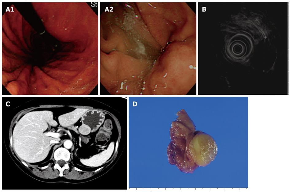
Figure 4 Endoscopic, EUS, abdominal computed tomography (CT), and gross findings of a schwannoma.
A: Endoscopic view of an ovoid subepithelial mass with a significant interval change; B: EUS shows a round, homogeneous, hypoechoic mass in the third gastric wall layer; C: CT scan shows a 2.8 cm × 2.2 cm homogeneous, well-defined, soft tissue mass on the upper body of the stomach; D: Gross findings of wedge resection reveal a 3 cm × 2.5 cm well-demarcated, round, firm, yellow mass.
- Citation: Lim YJ, Son HJ, Lee JS, Byun YH, Suh HJ, Rhee PL, Kim JJ, Rhee JC. Clinical course of subepithelial lesions detected on upper gastrointestinal endoscopy. World J Gastroenterol 2010; 16(4): 439-444
- URL: https://www.wjgnet.com/1007-9327/full/v16/i4/439.htm
- DOI: https://dx.doi.org/10.3748/wjg.v16.i4.439


