Copyright
copy;2010 Baishideng Publishing Group Co.
World J Gastroenterol. Oct 21, 2010; 16(39): 4986-4991
Published online Oct 21, 2010. doi: 10.3748/wjg.v16.i39.4986
Published online Oct 21, 2010. doi: 10.3748/wjg.v16.i39.4986
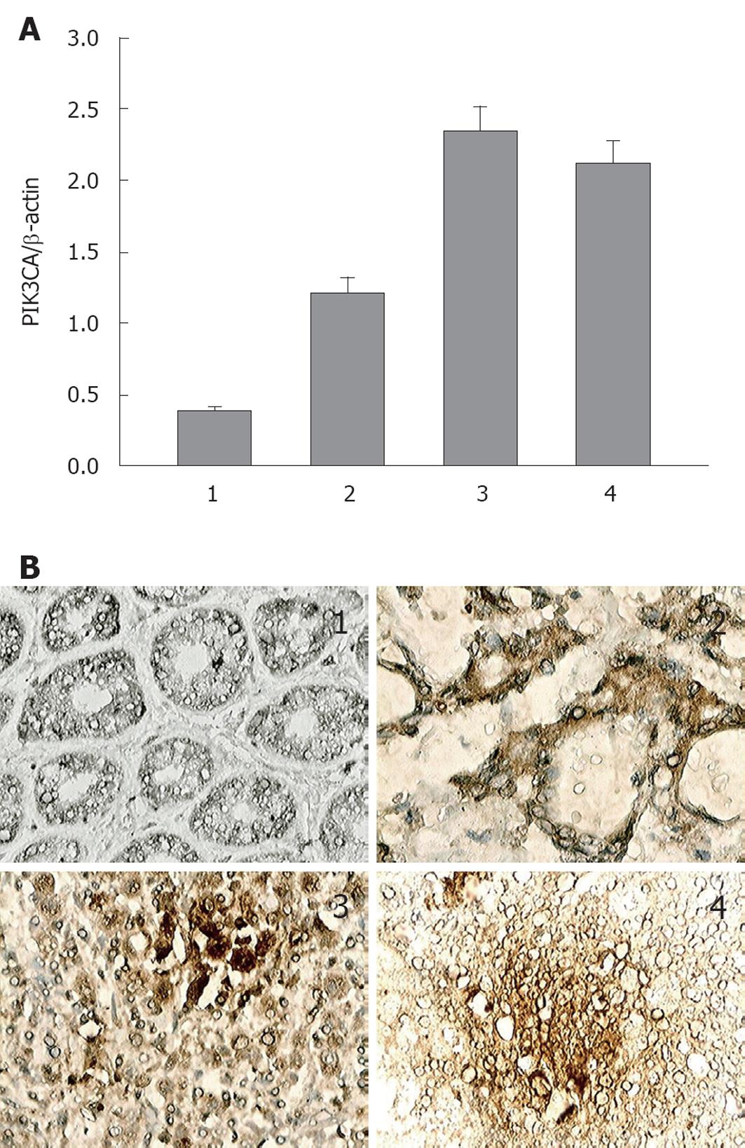
Figure 1 PIK3CA mRNA and protein expression in normal and gastric cancer tissues.
A: Expression analyses of PIK3CA mRNA determined by real-time quantitative polymerase chain reaction, values are shown as mean ± SD; B: Immunohistochemical study of PIK3CA (original magnification: × 400). Negative or weak expression of PIK3CA in normal tissues around tumor; Positive expression of PIK3CA in primary gastric cancer tissues. Strong positive expression of PIK3CA was detected in lymph node metastasis and distant metastasis gastric cancer tissues. 1: Normal gastric mucosa; 2: Primary gastric cancer; 3: Lymph node metastasis in gastric cancer; 4: Distant metastasis in gastric cancer.
Figure 2 Western blotting analyses of Akt phosphorylation.
A: Protein from the indicated tissues was Western blotting with anti-phospho-Akt (Ser473) and anti-Akt to analyze the effect on activation of phosphatidylinositol 3-kinase (PI3K)/Akt signaling. β-actin was used as a loading control; B: Relative expression level of p-Akt (Ser473) protein quantified by grey analysis; C: Relative expression level of total Akt protein quantified by grey analysis. p-AKT, phosphorylated Akt; Lane 1: Normal gastric mucosa; Lane 2: Primary gastric cancer; Lane 3: Lymph node metastasis in gastric cancer; Lane 4: Distant metastasis in gastric cancer. a,b,cSignificant difference was indicated with different lower-case letters (n = 3).
- Citation: Liu JF, Zhou XK, Chen JH, Yi G, Chen HG, Ba MC, Lin SQ, Qi YC. Up-regulation of PIK3CA promotes metastasis in gastric carcinoma. World J Gastroenterol 2010; 16(39): 4986-4991
- URL: https://www.wjgnet.com/1007-9327/full/v16/i39/4986.htm
- DOI: https://dx.doi.org/10.3748/wjg.v16.i39.4986


