Copyright
©2010 Baishideng Publishing Group Co.
World J Gastroenterol. Sep 21, 2010; 16(35): 4483-4490
Published online Sep 21, 2010. doi: 10.3748/wjg.v16.i35.4483
Published online Sep 21, 2010. doi: 10.3748/wjg.v16.i35.4483
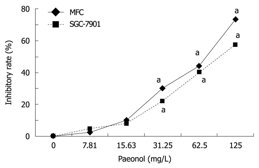
Figure 1 Paeonol inhibits the proliferation of mouse forestomach carcinoma and SGC-7901 cells in vitro.
Mouse forestomach carcinoma (MFC) or SGC-7901 cells (1 × 10⁴ cells) were incubated either alone (control) or in the presence of different concentrations of paeonol for 48 h and cell proliferation was measured by the 3-(4,5-dimethylthiazol-2-yl)-2,5-diphenyltetrazolium bromide method. Treatment with paeonol caused dose-dependent inhibition of cell proliferation. aP < 0.05, paeonol treated vs untreated control. Data are mean ± SD (n = 6).
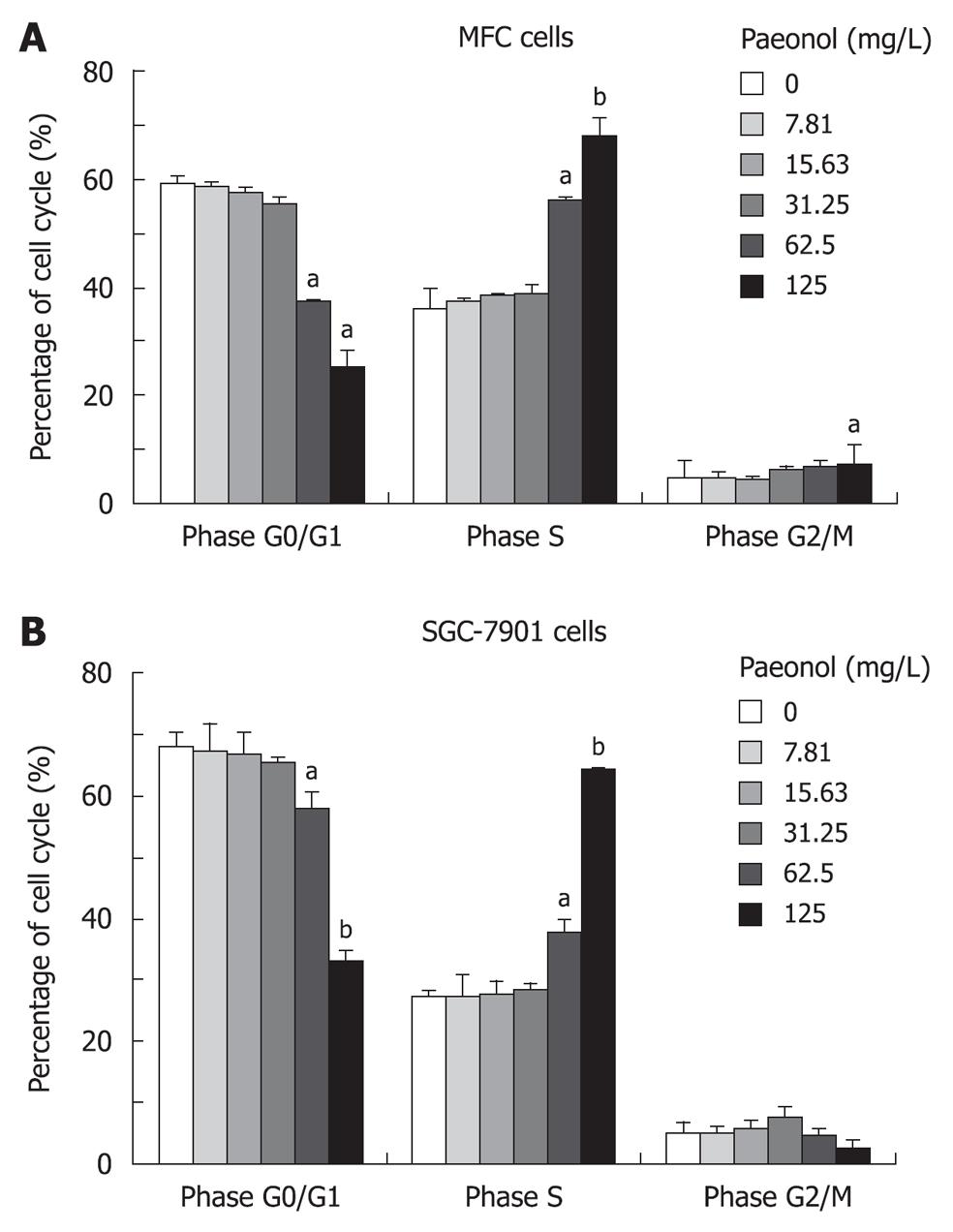
Figure 2 Effects of paeonol on cell cycle of mouse forestomach carcinoma and SGC-7901 cells in vitro.
A: Mouse forestomach carcinoma (MFC) cells; B: SGC-7901 cells. The number of cells in G0/G1, S, G2/M phases was determined by flow cytometry. Values are expressed as the mean ± SD from three replicate experiments (n = 6). aP < 0.05, bP < 0.01, paeonol treated vs untreated control.
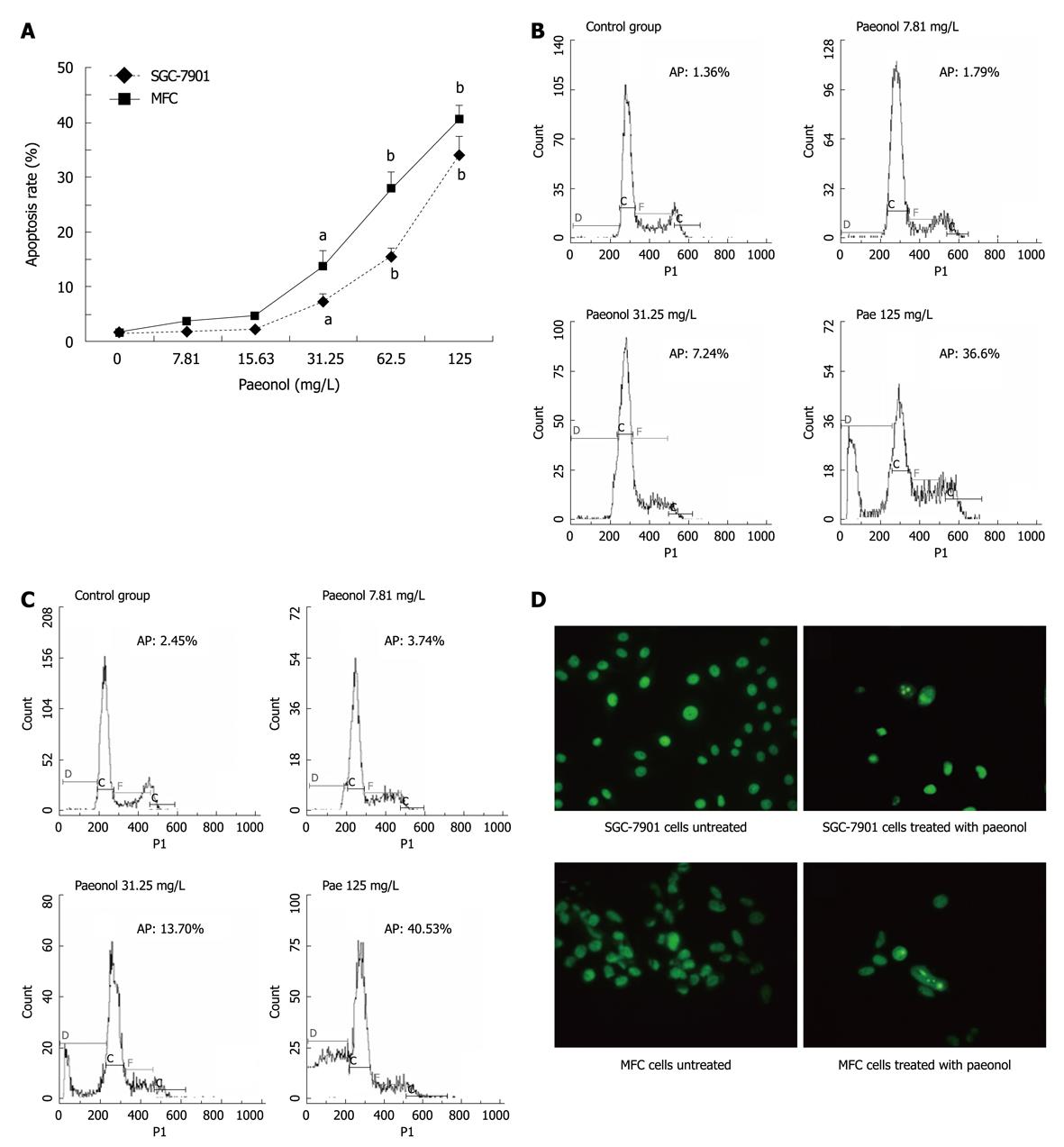
Figure 3 Induction of apoptosis by paeonol in cultured mouse forestomach carcinoma and SGC-7901 cells.
A: Paeonol increased the rate of apoptosis, as determined by the number of cells with sub-G1 DNA content by flow cytometry. Values are expressed as the mean ± SD from three replicate experiments. aP < 0.05, bP < 0.01, paeonol treated vs untreated control; B, C: Effects of paeonol on apoptosis of gastric cancer cell line SGC-7901 (B) and mouse forestomach carcinoma (MFC) cells (C) by flow cytometry. One representative flow cytometry tracing was shown from three replicate experiments. AP: Apoptotic percentage; D: Fluorescent staining of nuclei (× 320) in paeonol-treated and untreated cells by acridine orange. SGC-7901 and MFC cells treated with 62.5 mg/L paeonol for 48 h. Condensed and fragmented nuclei and apoptotic bodies were seen in the paeonol-treated cells, but not in the control.
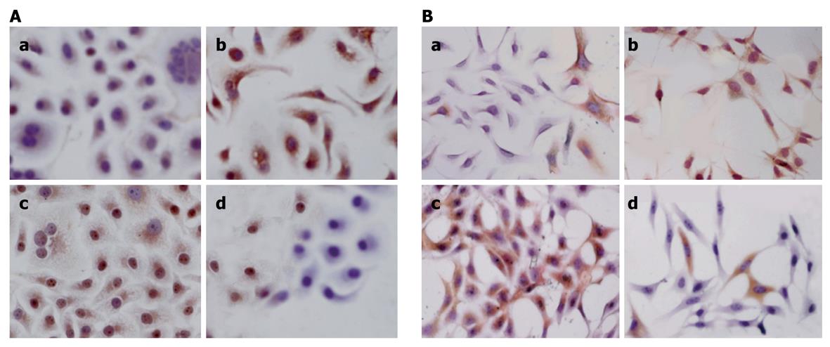
Figure 4 Expression of Bcl-2 and Bax in mouse forestomach carcinoma and SGC-7901 cells with or without paeonol treatment (× 200).
A: SGC-7901, cells were treated with 62.5 mg/L paeonol or with vehicle control for 48 h. a: Untreated control; b: Paeonol treatment. Bax expression was detected by immunohistochemical analysis. Cells treated with paeonol had a significant increase in the expression of Bax. c: Untreated control; d: Paeonol treatment. Bcl-2 expression was detected by immunohistochemical analysis. Cells treated with paeonol had a significant decrease in the expression of Bcl-2; B: Mouse forestomach carcinoma, cells were treated with 62.5 mg/L paeonol or with vehicle control for 48 h. a: Untreated control; b: Paeonol treatment. Bax expression was detected by immunohistochemical analysis. Cells treated with paeonol had a significant increase in the expression of Bax. c: Untreated control; d: Paeonol treatment. Bcl-2 expression was detected by immunohistochemical analysis. Cells treated with paeonol had a significant decrease in the expression of Bcl-2.
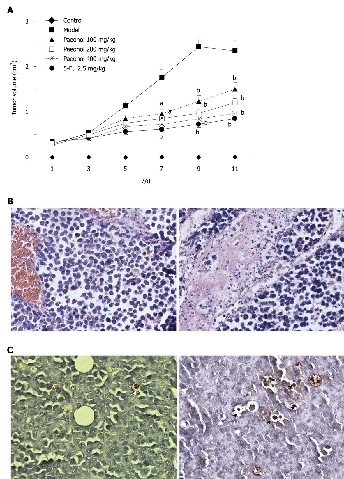
Figure 5 Paeonol inhibits gastric tumor growth in vivo associated with induction of apoptosis and inhibiting proliferation.
A: Effects of paeonol or 5-fluorouracil (5-FU) in tumor volume. Values are expressed as the mean ± SD (n = 12-14). aP < 0.05, bP < 0.01, paeonol treated vs model group; B: Hematoxylin-eosin-stained sections of mouse forestomach carcinoma (MFC) tumors from untreated controls showed numerous cells in mitosis (left), paeonol (200 mg/kg) treatment suppressed cell proliferation, as indicated by the small number of mitotic cells (right) (× 400); C: TUNEL stained sections of MFC tumors from untreated control displayed occasionally apoptotic cells (left). Tumors from mice treated with paeonol (200 mg/kg) had a prominent population of cells with condensed or fragmented nuclei and cytoplasm shrinkage, indicating apoptosis (right) (× 400). The arrows indicate apoptotic cells.
-
Citation: Li N, Fan LL, Sun GP, Wan XA, Wang ZG, Wu Q, Wang H. Paeonol inhibits tumor growth in gastric cancer
in vitro andin vivo . World J Gastroenterol 2010; 16(35): 4483-4490 - URL: https://www.wjgnet.com/1007-9327/full/v16/i35/4483.htm
- DOI: https://dx.doi.org/10.3748/wjg.v16.i35.4483


