Copyright
©2010 Baishideng.
World J Gastroenterol. Jul 21, 2010; 16(27): 3472-3474
Published online Jul 21, 2010. doi: 10.3748/wjg.v16.i27.3472
Published online Jul 21, 2010. doi: 10.3748/wjg.v16.i27.3472
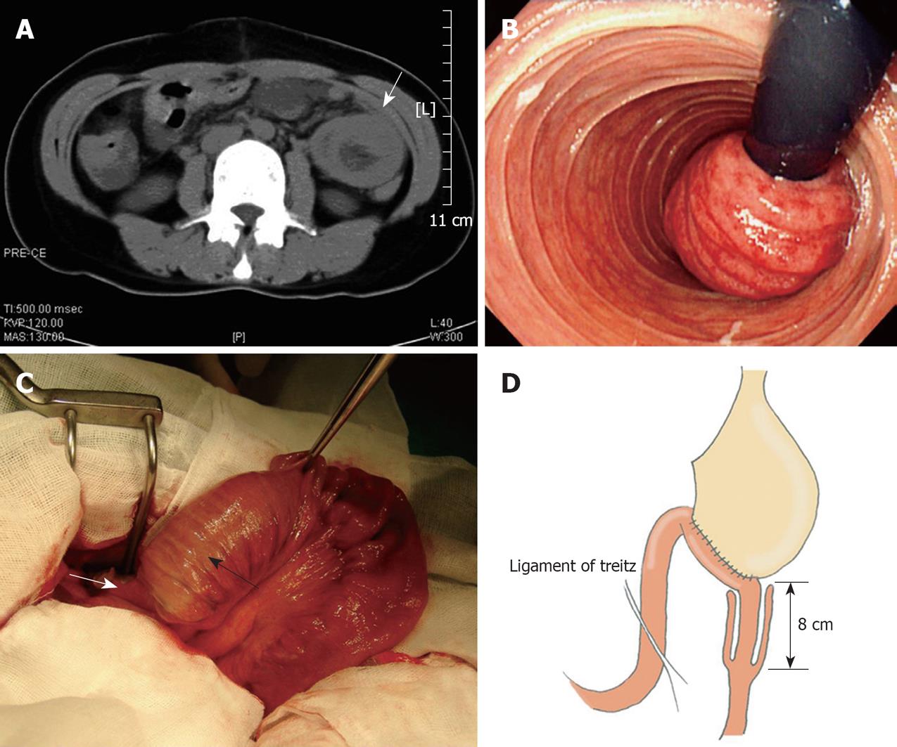
Figure 1 Radiologic, endoscopic and intraoperative findings of anterograde jejunojejunal intussusception.
A: Abdominal computed tomography showed a non-homogeneous mass (arrow) in the left upper quadrant of the abdomen; B: A J-turn view of gastrofibroscopic examination demonstrated a congested jejunal mass (intussusceptum); C: Intraoperative findings revealed that the intussusception started just below the gastrojejunostomy anastomosis. The black arrow indicates the intussuscipiens, and the white arrow indicates the intussusceptum; D: Schematic diagram of the intussusception at the efferent-loop.
- Citation: Kwak JM, Kim J, Suh SO. Anterograde jejunojejunal intussusception resulted in acute efferent loop syndrome after subtotal gastrectomy. World J Gastroenterol 2010; 16(27): 3472-3474
- URL: https://www.wjgnet.com/1007-9327/full/v16/i27/3472.htm
- DOI: https://dx.doi.org/10.3748/wjg.v16.i27.3472


