Copyright
©2010 Baishideng.
World J Gastroenterol. Jun 21, 2010; 16(23): 2841-2850
Published online Jun 21, 2010. doi: 10.3748/wjg.v16.i23.2841
Published online Jun 21, 2010. doi: 10.3748/wjg.v16.i23.2841
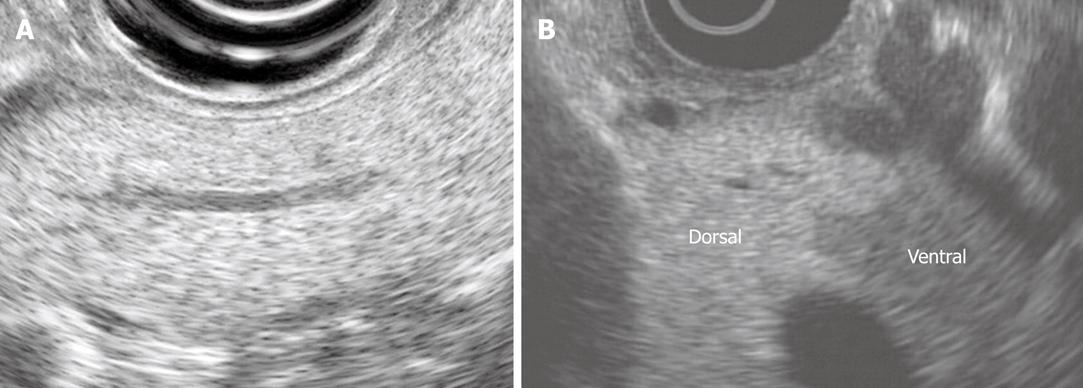
Figure 1 The normal endosonographic appearance of the pancreas.
A: View of pancreatic body from gastric station. The parenchyma is homogeneous and granular (“salt and pepper”). The duct is neither dilated nor ectatic; B: Dorsal ventral anlage. The ventral pancreas is relatively echogenic compared with the dorsal pancreas. There is a distinct border between the dorsal and ventral pancreas.
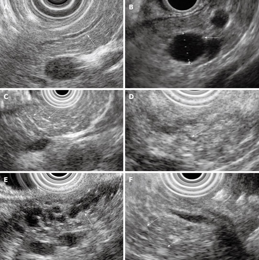
Figure 2 Examples of endoscopic ultrasound (EUS) chronic pancreatitis (CP) criteria.
A: Hyperechoic duct wall (arrow); B: Cyst (arrow); C: Hyperechoic strands (arrows); D: Visible side-branch (arrow); E: Dilated and irregular main pancreatic duct with visible side-branches (arrow); F: Hyperechoic foci (arrows).
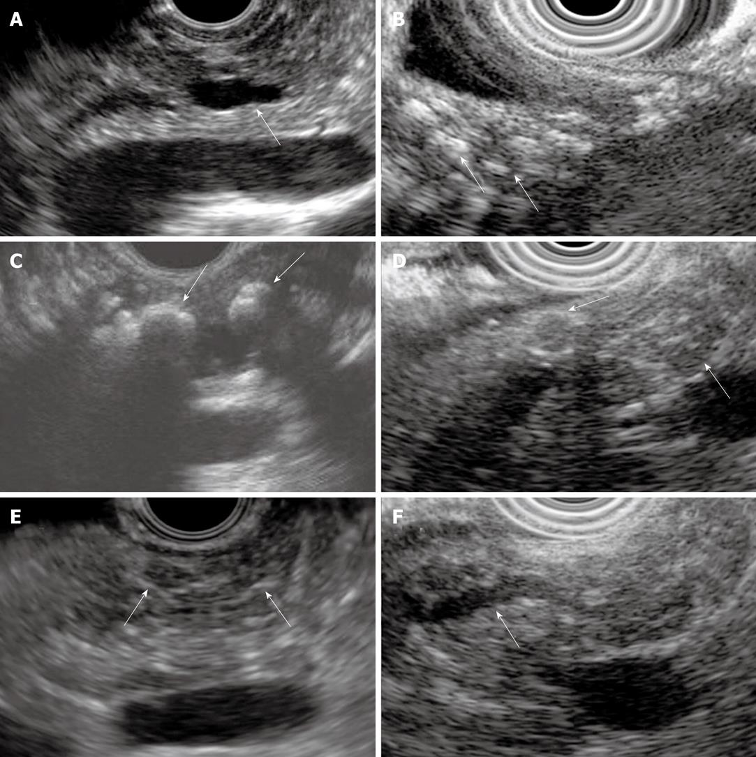
Figure 3 Examples of EUS CP criteria.
A: Dilated main pancreatic duct (arrow); B: Parenchymal calcifications (arrows); C: Main duct calcifications (arrows); D: Lobules (arrows); E: Stranding (arrows); F: Irregular main pancreatic duct (arrow).
- Citation: Stevens T, Parsi MA. Endoscopic ultrasound for the diagnosis of chronic pancreatitis. World J Gastroenterol 2010; 16(23): 2841-2850
- URL: https://www.wjgnet.com/1007-9327/full/v16/i23/2841.htm
- DOI: https://dx.doi.org/10.3748/wjg.v16.i23.2841


