Copyright
©2010 Baishideng.
World J Gastroenterol. May 28, 2010; 16(20): 2566-2570
Published online May 28, 2010. doi: 10.3748/wjg.v16.i20.2566
Published online May 28, 2010. doi: 10.3748/wjg.v16.i20.2566
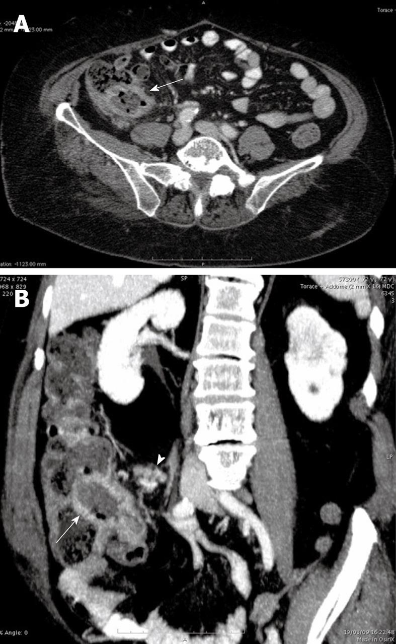
Figure 1 The axial computed tomography (CT) image (A) and the multiplanar reformatted (MPR) coronal oblique image (B) showed thickened walls of the terminal ileum with mild pseudoaneurysmal dilatation of the lumen (arrows) and multiple ileo-cecal lymph nodes (arrow head).
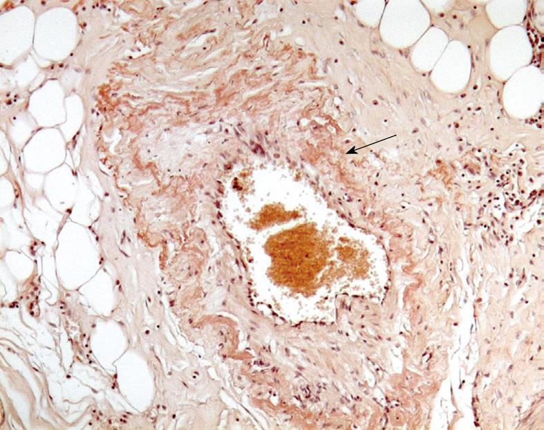
Figure 2 Intestinal amyloid (“congophilic”) angiopathy.
Amyloid in the intramural vessels of terminal ileum walls (arrow) takes the Congo red stains (× 270).
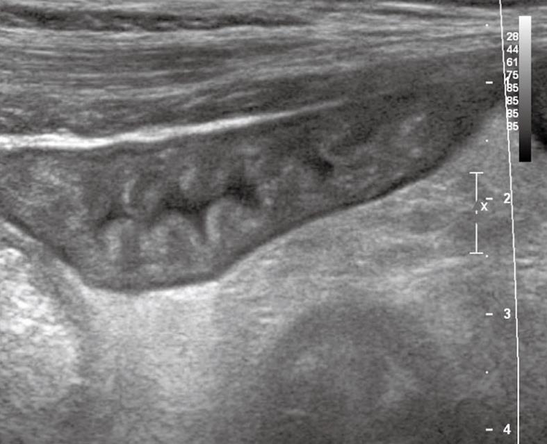
Figure 3 Ultrasound images.
Thickened and stratified bowel walls with hypertrophy of the mucosal and sub-mucosal strata and increased number of folds due to amyloid deposition.
Figure 4 MRI changes of the small bowel.
A and B: Coronal T2-weighted half-Fourier single-shot turbo spin-echo magnetic resonance images showed diffuse intestinal wall symmetrical thickening with increased number and hypertrophy of the folds (A, arrow) and “jejunalization” of the ileum (B, arrow); C: 3D T1-fast imaging in the steady-state precession after iv administration of contrast agent showed diffuse enhancement of intestinal walls (arrows).
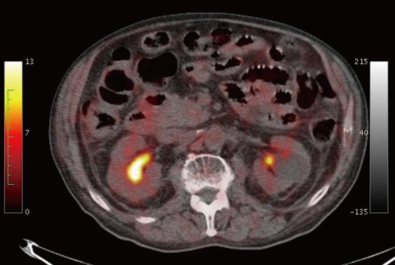
Figure 5 18FDG-positron emission tomography (PET)/CT images did not show focal or diffuse increase of the tracer uptake of small bowel walls.
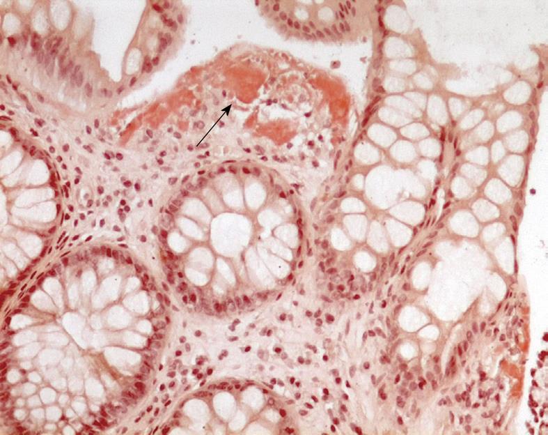
Figure 6 Colonic biopsy.
Submucosal amyloid deposition (arrow) (Congo red stains, × 270).
- Citation: Mainenti PP, Segreto S, Mancini M, Rispo A, Cozzolino I, Masone S, Rinaldi CR, Nardone G, Salvatore M. Intestinal amyloidosis: Two cases with different patterns of clinical and imaging presentation. World J Gastroenterol 2010; 16(20): 2566-2570
- URL: https://www.wjgnet.com/1007-9327/full/v16/i20/2566.htm
- DOI: https://dx.doi.org/10.3748/wjg.v16.i20.2566


