Copyright
©2010 Baishideng.
World J Gastroenterol. May 28, 2010; 16(20): 2496-2503
Published online May 28, 2010. doi: 10.3748/wjg.v16.i20.2496
Published online May 28, 2010. doi: 10.3748/wjg.v16.i20.2496
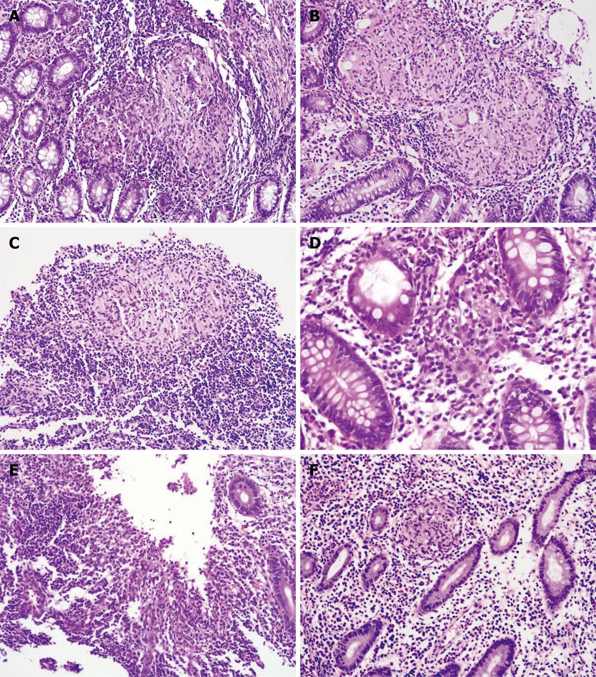
Figure 1 Histopathological parameters of intestinal tuberculosis (ITB) and Crohn’s disease (CD).
A: Confluent granuloma in ITB; B: Confluent granuloma with caseation necrosis and Langhans giant cells in ITB; C: Granuloma with lymphoid cuff in ITB; D: Vague granuloma in CD; E: Band of epithelioid histiocytes in ulcer base in ITB; F: Small granuloma in CD.
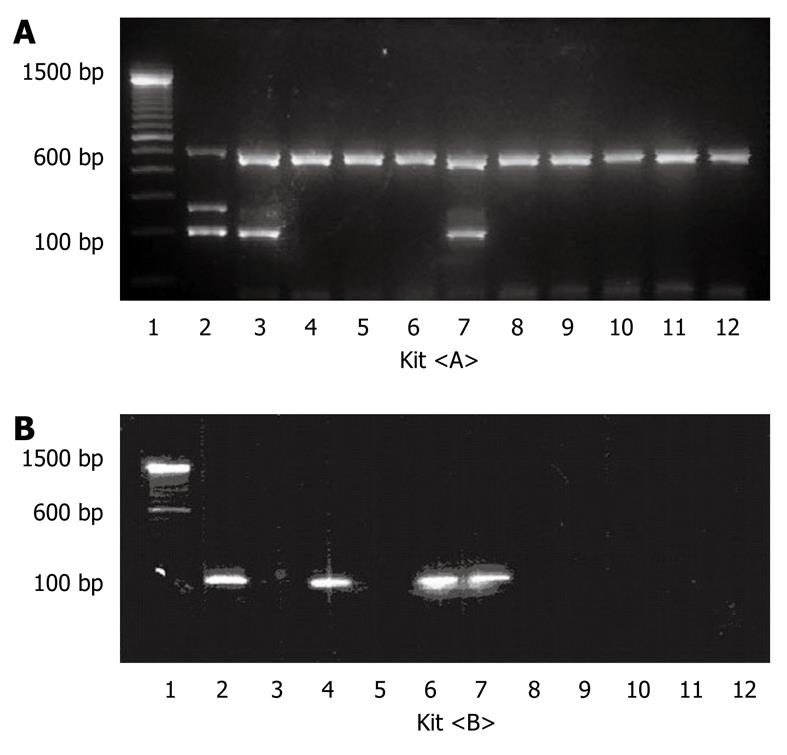
Figure 2 Polymerase chain reaction (PCR) product by kit <A> and kit <B>.
A: Number 3 and 7 showing positive result by kit <A>; B: Number 2, 4, 6, 7 showing positive result by kit <B>.
- Citation: Jin XJ, Kim JM, Kim HK, Kim L, Choi SJ, Park IS, Han JY, Chu YC, Song JY, Kwon KS, Kim EJ. Histopathology and TB-PCR kit analysis in differentiating the diagnosis of intestinal tuberculosis and Crohn’s disease. World J Gastroenterol 2010; 16(20): 2496-2503
- URL: https://www.wjgnet.com/1007-9327/full/v16/i20/2496.htm
- DOI: https://dx.doi.org/10.3748/wjg.v16.i20.2496


