Copyright
©2010 Baishideng.
World J Gastroenterol. May 14, 2010; 16(18): 2314-2316
Published online May 14, 2010. doi: 10.3748/wjg.v16.i18.2314
Published online May 14, 2010. doi: 10.3748/wjg.v16.i18.2314
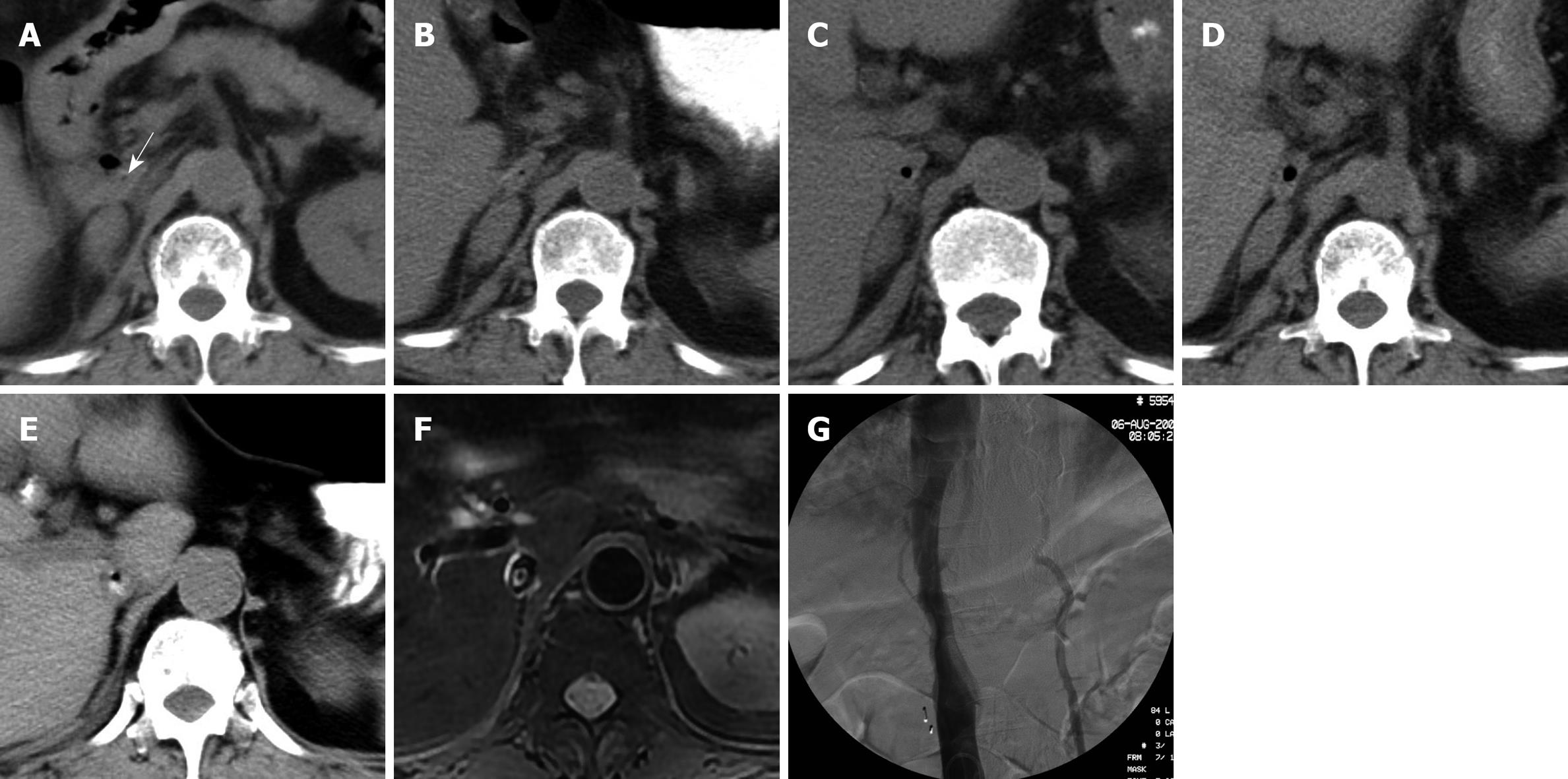
Figure 1 During the 2-mo hospital stay, a small, low-density air bubble appeared in the inferior vena cava (IVC) and gradually enlarged on follow-up computed tomography (CT) scans.
Enteric contrast medium leaked into the IVC. Magnetic resonance imaging (MRI) clearly demonstrated the high-signal enteric contrast medium or thrombus and signal-void air bubbles. Cavography did not reveal the thrombus in the IVC. A: CT scan on the second hospital day. A low-density dot loomed up in the IVC (arrow). A mass in the right side adrenal gland was also seen; B: CT scan on the 19th hospital day. The low-density dot became more evident; C, D: CT scan on the 59th and 67th hospital day. The low-density dot (air bubble) was gradually enlarged; E: CT scan on the 73rd hospital day with water-soluble enteric contrast demonstrated air bubble surrounded by contrast medium in the IVC; F: MRI T2WI on the 74th hospital day demonstrated the high signal enteric contrast medium or thrombus and signal void air bubble in the IVC; G: Inferior vena cavogram on the 75th hospital day showed no evident thrombus in the IVC.
- Citation: Guo Y, Zhang YQ, Lin W. Radiological diagnosis of duodenocaval fistula: A case report and literature review. World J Gastroenterol 2010; 16(18): 2314-2316
- URL: https://www.wjgnet.com/1007-9327/full/v16/i18/2314.htm
- DOI: https://dx.doi.org/10.3748/wjg.v16.i18.2314


