Copyright
©2010 Baishideng.
World J Gastroenterol. Apr 28, 2010; 16(16): 2061-2064
Published online Apr 28, 2010. doi: 10.3748/wjg.v16.i16.2061
Published online Apr 28, 2010. doi: 10.3748/wjg.v16.i16.2061
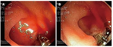
Figure 1 Endoscopic images.
A: The bleeding point is pinched with a hemostatic forceps; B: Coagulation delivery at the retracted bleeding point.
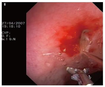
Figure 2 An opened hemostatic forceps while washing out the blood after coagulation.
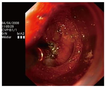
Figure 3 Coagulating the bleeding vessels during cap-assisted endoscopic mucosal resection (EMR).
- Citation: Coumaros D, Tsesmeli N. Active gastrointestinal bleeding: Use of hemostatic forceps beyond endoscopic submucosal dissection. World J Gastroenterol 2010; 16(16): 2061-2064
- URL: https://www.wjgnet.com/1007-9327/full/v16/i16/2061.htm
- DOI: https://dx.doi.org/10.3748/wjg.v16.i16.2061


