Copyright
©2010 Baishideng.
World J Gastroenterol. Apr 28, 2010; 16(16): 2023-2027
Published online Apr 28, 2010. doi: 10.3748/wjg.v16.i16.2023
Published online Apr 28, 2010. doi: 10.3748/wjg.v16.i16.2023
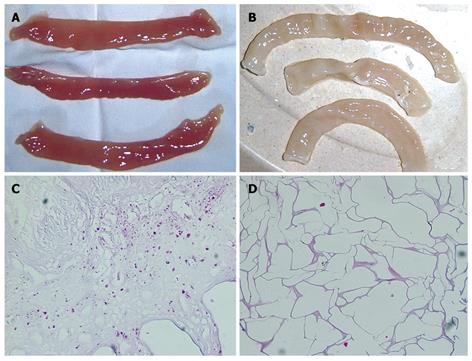
Figure 1 Acellular dermal matrix.
A: Segments of the small intestine; B: Segments of acellular dermal matrix; C, D: Hematoxylin and eosin–stained histological photomicrography showed that all the cellular elements of the small intestine have been removed (C: × 100, D: × 400)
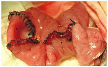
Figure 2 Surgical procedures.
a: ADM; arrow: Side-to-side anastomosis.
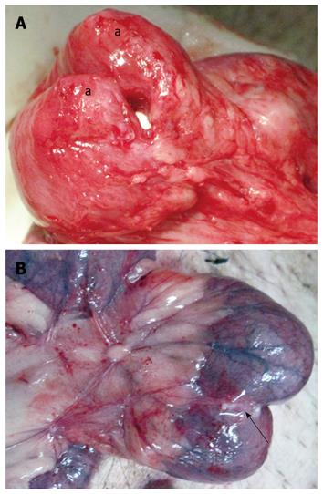
Figure 3 Postoperative gross specimen.
A: ADM has been absorbed completely (a: Blind-ended pouch); B: The graft was shrinking (arrow: The ADM material).
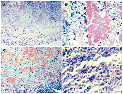
Figure 4 Histological photomicrography of postoperative ADM.
Focal fibrinoid necroses and infiltration of a large amount of neutrophils and leukomonocytes in the first week (HE stain, A: × 100, B: × 400); a further development of the inflammation in the second week (HE stain, C: × 100, D: × 400).
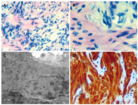
Figure 5 Structures of the ADM were contorted in the narrow-ring.
A: × 100; B: × 200; C: Transmission electron microscopy, × 20 000; a: Fibroblast; D: Immunocytochemical stain with smooth muscle actin showed smooth muscle cells in the narrow-ring.
- Citation: Xu HM, Wang ZJ, Han JG, Ma HC, Zhao B, Zhao BC. Application of acellular dermal matrix for intestinal elongation in animal models. World J Gastroenterol 2010; 16(16): 2023-2027
- URL: https://www.wjgnet.com/1007-9327/full/v16/i16/2023.htm
- DOI: https://dx.doi.org/10.3748/wjg.v16.i16.2023


