Copyright
©2010 Baishideng.
World J Gastroenterol. Mar 28, 2010; 16(12): 1522-1526
Published online Mar 28, 2010. doi: 10.3748/wjg.v16.i12.1522
Published online Mar 28, 2010. doi: 10.3748/wjg.v16.i12.1522
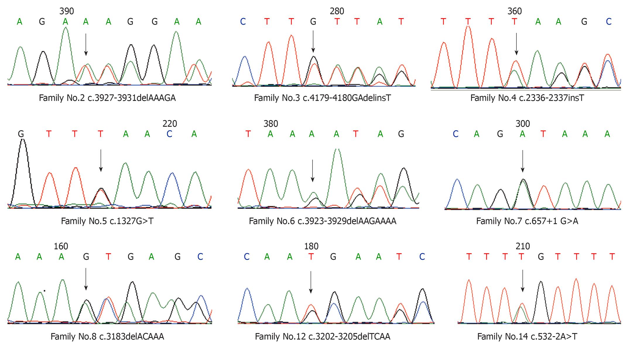
Figure 1 DNA sequencing of micromutations.
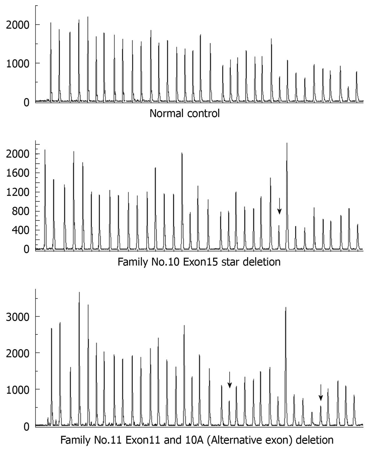
Figure 2 Peak of large fragment deletion detected by multiplex ligation-dependent probe amplification (MLPA).
Arrows show the reduced relative peak area of the amplification product of that probe which means heterozygous deletions of corresponding exons.
-
Citation: Sheng JQ, Cui WJ, Fu L, Jin P, Han Y, Li SJ, Fan RY, Li AQ, Zhang MZ, Li SR.
APC gene mutations in Chinese familial adenomatous polyposis patients. World J Gastroenterol 2010; 16(12): 1522-1526 - URL: https://www.wjgnet.com/1007-9327/full/v16/i12/1522.htm
- DOI: https://dx.doi.org/10.3748/wjg.v16.i12.1522


