Copyright
©2010 Baishideng.
World J Gastroenterol. Mar 14, 2010; 16(10): 1296-1298
Published online Mar 14, 2010. doi: 10.3748/wjg.v16.i10.1296
Published online Mar 14, 2010. doi: 10.3748/wjg.v16.i10.1296
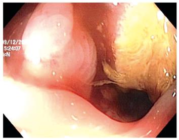
Figure 1 Colonoscopic appearance of an ulcer at the hepatic flexure.
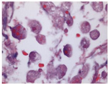
Figure 2 Colonoscopic biopsy showing amoeba (× 40).
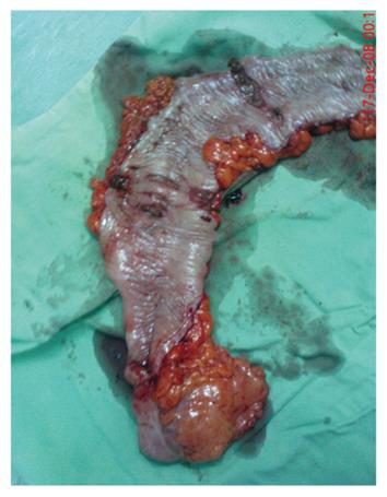
Figure 3 Gross appearance of ulcers in the colectomy specimen.
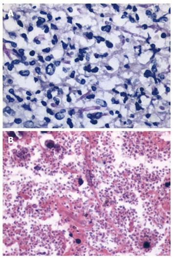
Figure 4 Histopathological examination.
A: Mycelial formations (Periodic acid-Schiff, × 40); B: Yeast formations (HE, × 40).
- Citation: Koh PS, Roslani AC, Vimal KV, Shariman M, Umasangar R, Lewellyn R. Concurrent amoebic and histoplasma colitis: A rare cause of massive lower gastrointestinal bleeding. World J Gastroenterol 2010; 16(10): 1296-1298
- URL: https://www.wjgnet.com/1007-9327/full/v16/i10/1296.htm
- DOI: https://dx.doi.org/10.3748/wjg.v16.i10.1296


