Copyright
©2010 Baishideng.
World J Gastroenterol. Mar 14, 2010; 16(10): 1293-1295
Published online Mar 14, 2010. doi: 10.3748/wjg.v16.i10.1293
Published online Mar 14, 2010. doi: 10.3748/wjg.v16.i10.1293
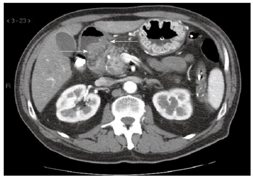
Figure 1 Contrast-enhanced CT of upper abdomen demonstrating a duodenal mass which narrowed the lumen (arrows).
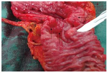
Figure 2 Duodenum.
Arrows indicate the EP adenocarcinoma into the duodenal wall; the scalpel indicates the Ampulla of Vater.
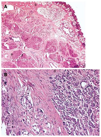
Figure 3 Histological findings.
A: Duodenal wall (a) and ectopic pancreatic tissue (b) (HE, × 25); B: Adenocarcinoma arising from ectopic pancreatic tissue (c) and ectopic pancreatic tissue (d) (HE, × 200).
- Citation: Bini R, Voghera P, Tapparo A, Nunziata R, Demarchi A, Capocefalo M, Leli R. Malignant transformation of ectopic pancreatic cells in the duodenal wall. World J Gastroenterol 2010; 16(10): 1293-1295
- URL: https://www.wjgnet.com/1007-9327/full/v16/i10/1293.htm
- DOI: https://dx.doi.org/10.3748/wjg.v16.i10.1293


