Copyright
©2009 The WJG Press and Baishideng.
World J Gastroenterol. Nov 21, 2009; 15(43): 5397-5408
Published online Nov 21, 2009. doi: 10.3748/wjg.15.5397
Published online Nov 21, 2009. doi: 10.3748/wjg.15.5397
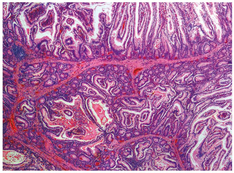
Figure 1 Hamartoma, a typical PJS polyp demonstrating the arborizing pattern of smooth-muscle proliferation.
HE staining, magnification 100 ×. (Courtesy of Professor A. Ryska, MD, PhD).
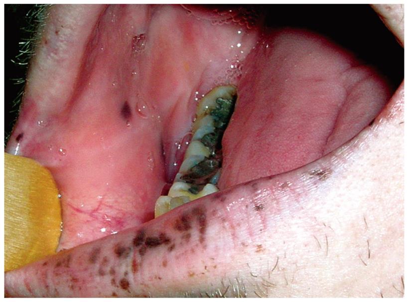
Figure 2 Pigmentations of the lips and oral mucosa.
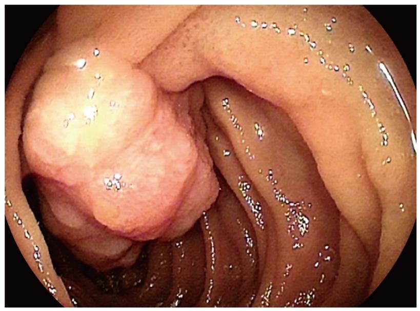
Figure 3 Hamartoma of the jejunum.
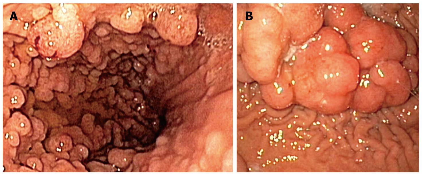
Figure 4 Hamartomas of the stomach.
A: Multiple; B: Voluminous.
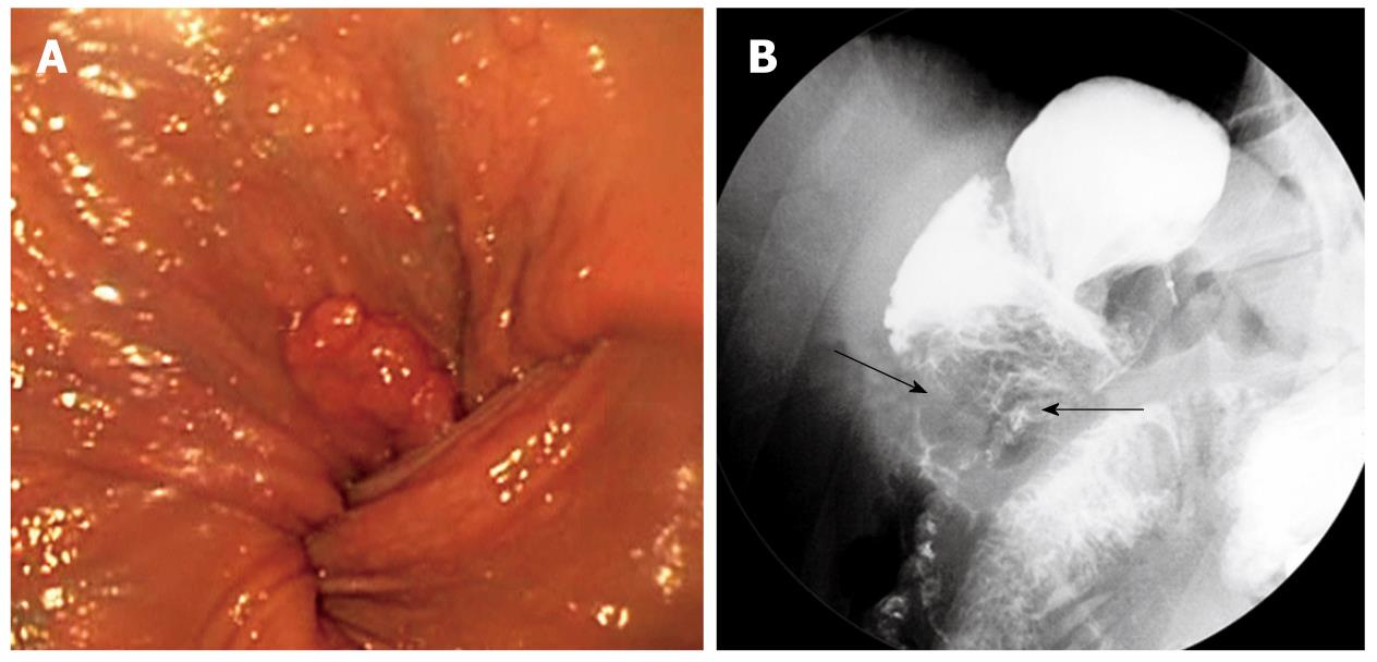
Figure 5 Gastric outlet obstruction (same patient as seen in Figure 4B).
A: Endoscopic view; B: fluoroscopy, mass of polyps marked by arrows.
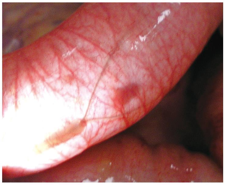
Figure 6 Intra-operative enteroscopy.
Polypectomy snare over a small polyp shines through the intestinal wall.
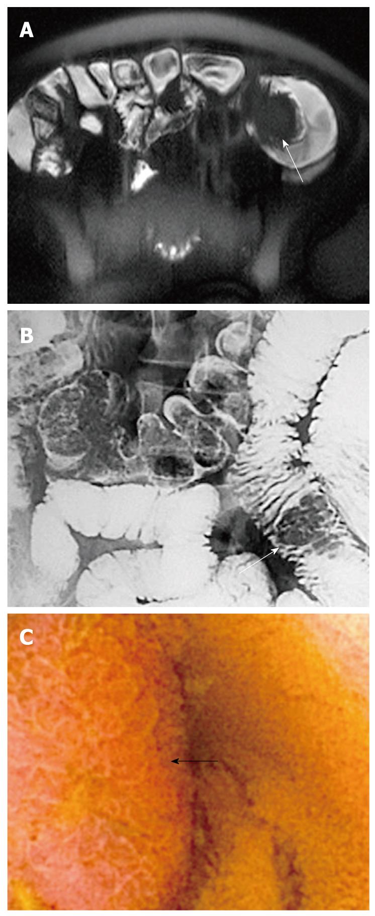
Figure 7 Hamartoma of the jejunum (arrow).
A: Magnetic resonance imaging; B: Enteroclysis; C: Capsule enteroscopy.
- Citation: Kopacova M, Tacheci I, Rejchrt S, Bures J. Peutz-Jeghers syndrome: Diagnostic and therapeutic approach. World J Gastroenterol 2009; 15(43): 5397-5408
- URL: https://www.wjgnet.com/1007-9327/full/v15/i43/5397.htm
- DOI: https://dx.doi.org/10.3748/wjg.15.5397


