Copyright
©2009 The WJG Press and Baishideng.
World J Gastroenterol. Nov 7, 2009; 15(41): 5239-5241
Published online Nov 7, 2009. doi: 10.3748/wjg.15.5239
Published online Nov 7, 2009. doi: 10.3748/wjg.15.5239
Figure 1 CT scan in abdomen showing a mass tumor between left hepatic lobe and spleen directly involving the upper pole of spleen and almost making no invasion into the liver (A-D).
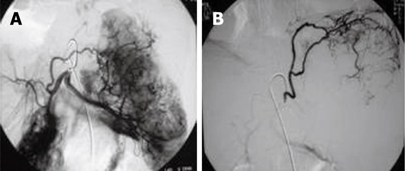
Figure 2 Celiac and hepatic arteriography confirmed the mass lesions taking blood from left hepatic artery and splenic artery (A) and inferior phrenic artery (B).
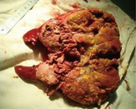
Figure 3 Postoperative photography showing the lesions directly involving the upper pole of spleen.
Figure 4 Histopathology showing the splenic metastasis of hepatocellular carcinoma (HE, × 40).
- Citation: Yan ML, Wang YD, Lai ZD, Tian YF, Chen HB, Qiu FN, Zhou SQ. Pedunculated hepatocellular carcinoma and splenic metastasis. World J Gastroenterol 2009; 15(41): 5239-5241
- URL: https://www.wjgnet.com/1007-9327/full/v15/i41/5239.htm
- DOI: https://dx.doi.org/10.3748/wjg.15.5239


