Copyright
©2009 The WJG Press and Baishideng.
World J Gastroenterol. Nov 7, 2009; 15(41): 5218-5220
Published online Nov 7, 2009. doi: 10.3748/wjg.15.5218
Published online Nov 7, 2009. doi: 10.3748/wjg.15.5218
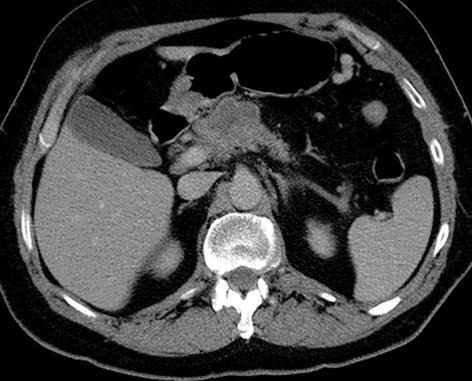
Figure 1 CT pneumocolon, which suggested the presence of advanced pancreatic malignancy.
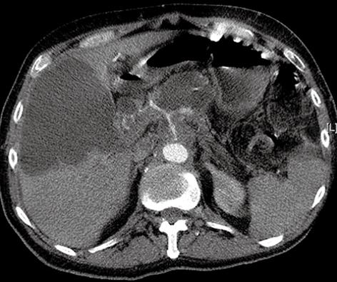
Figure 2 Large intrahepatic cystic lesion with bile duct compression caused by enlargement of the pancreatic neoplasm.
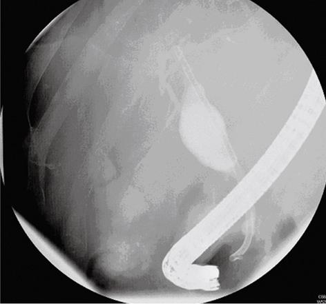
Figure 3 ERCP demonstrating a smooth stricture in the distal CBD, with dilated common hepatic ducts above.
Contrast material was seen to flow within the biloma collection, which implied communication with the intrahepatic ducts.
- Citation: Trivedi PJ, Gupta P, Phillips-Hughes J, Ellis A. Biloma: An unusual complication in a patient with pancreatic cancer. World J Gastroenterol 2009; 15(41): 5218-5220
- URL: https://www.wjgnet.com/1007-9327/full/v15/i41/5218.htm
- DOI: https://dx.doi.org/10.3748/wjg.15.5218


