Copyright
©2009 The WJG Press and Baishideng.
World J Gastroenterol. Oct 7, 2009; 15(37): 4729-4731
Published online Oct 7, 2009. doi: 10.3748/wjg.15.4729
Published online Oct 7, 2009. doi: 10.3748/wjg.15.4729
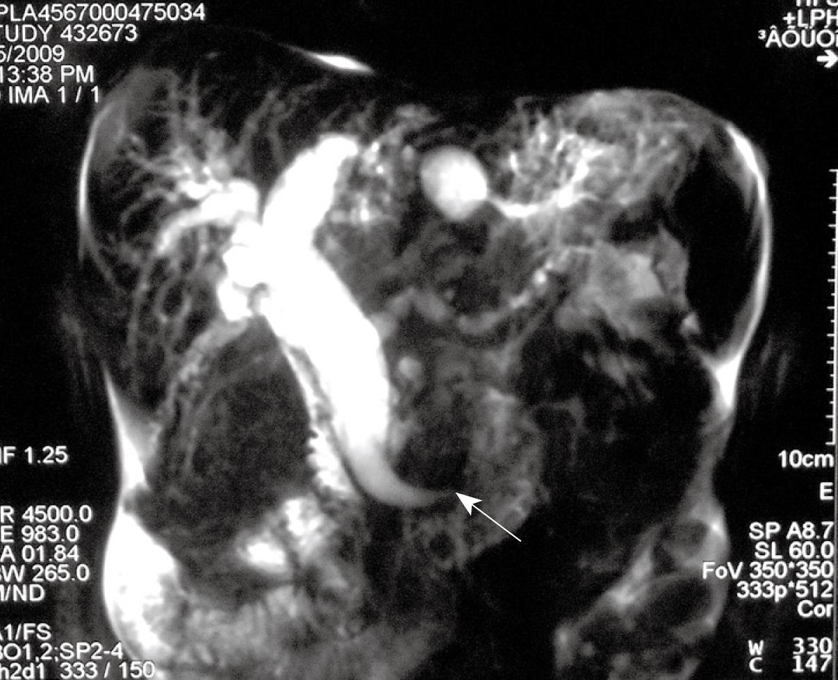
Figure 1 Magnetic resonance imaging showed the expansion of hepatic biliary duct and the ectopic hepatopancreatic ampulla (arrow) draining into the fourth part of the duodenum.
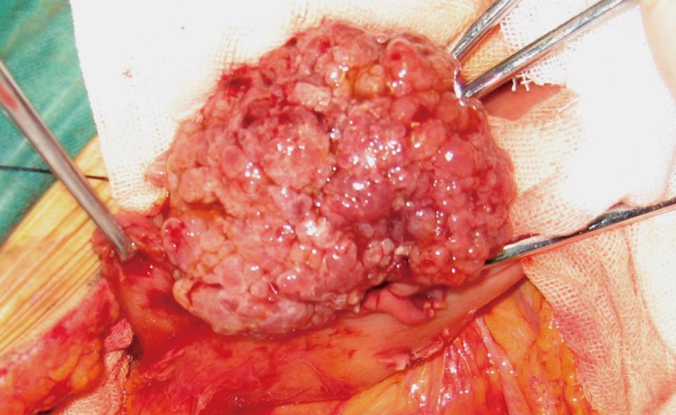
Figure 2 A soft texture tumor about 6 cm × 5 cm × 5 cm with a peduncle conjunct to the ampulla.
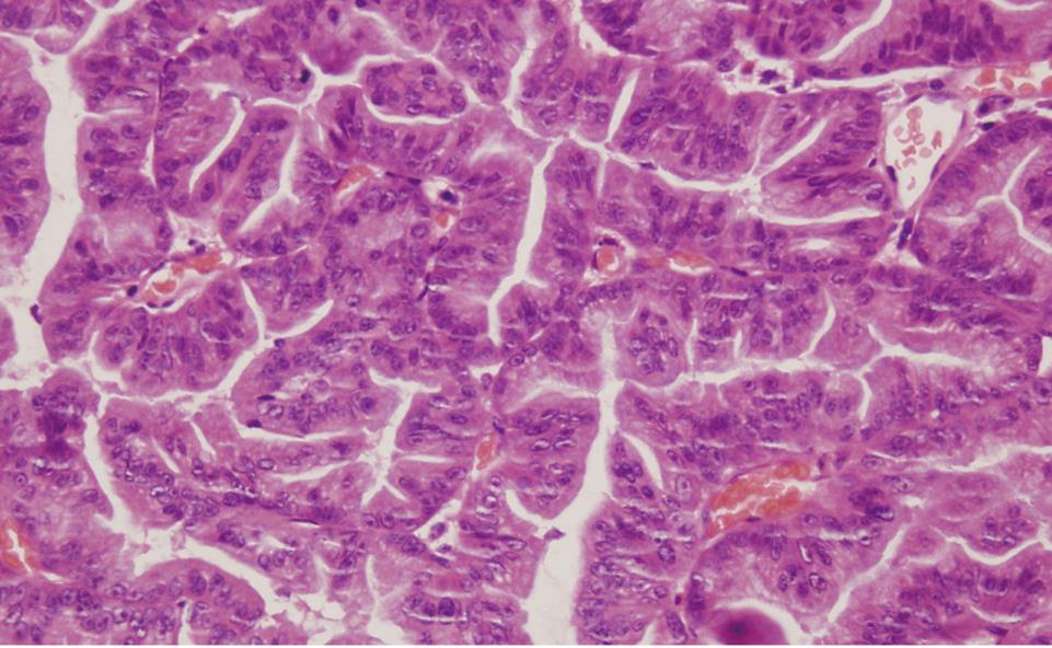
Figure 3 Microscopic examination (HE, × 400) showed severe dysplasia and focal carcinomatous change.
- Citation: Jin SG, Chen ZY, Yan LN, Zeng Y, Huang W, Xu N. A rare case of periampullary carcinoma with ectopic ending of Vater’s ampulla. World J Gastroenterol 2009; 15(37): 4729-4731
- URL: https://www.wjgnet.com/1007-9327/full/v15/i37/4729.htm
- DOI: https://dx.doi.org/10.3748/wjg.15.4729


