Copyright
©2009 The WJG Press and Baishideng.
World J Gastroenterol. Jul 21, 2009; 15(27): 3431-3433
Published online Jul 21, 2009. doi: 10.3748/wjg.15.3431
Published online Jul 21, 2009. doi: 10.3748/wjg.15.3431
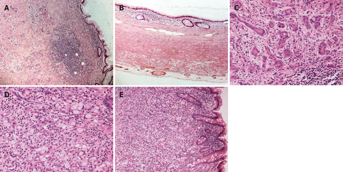
Figure 1 Histopathological examination.
A: Mucinous cystadenoma of the appendix combined with infiltrating goblet cell carcinoid (HE, × 50); B: The mucinous cystadenoma of the appendix showing lumen lined by mucous-containing bland epithelial cells (HE, × 100); C: Cells with neuroendocrine cytonuclear features in goblet cell carcinoid (HE, × 200); D: Mucin-filled neoplastic cells of goblet cell carcinoid (HE, × 200); E: The infiltrating nests of goblet cell carcinoid appear to arise from the basiglandular region of the intestinal crypts in close proximity to the mucinous cystadenoma (HE, × 100).
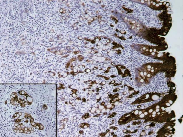
Figure 2 Immunohistochemically.
The neoplastic cells of the goblet cell carcinoid expressed strong and diffuse positivity for cytokeratin 20 (× 100).
- Citation: Alsaad KO, Serra S, Chetty R. Combined goblet cell carcinoid and mucinous cystadenoma of the vermiform appendix. World J Gastroenterol 2009; 15(27): 3431-3433
- URL: https://www.wjgnet.com/1007-9327/full/v15/i27/3431.htm
- DOI: https://dx.doi.org/10.3748/wjg.15.3431


