Copyright
©2009 The WJG Press and Baishideng.
World J Gastroenterol. May 14, 2009; 15(18): 2287-2289
Published online May 14, 2009. doi: 10.3748/wjg.15.2287
Published online May 14, 2009. doi: 10.3748/wjg.15.2287
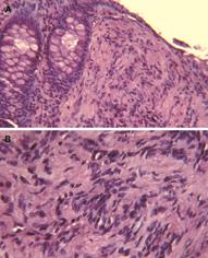
Figure 1 Histological features of the lesion.
Low- (A) and high (B)-power magnification of haematoxylin and eosin stained tissue sections of colorectal mucosa. A diffuse, Schwann cell proliferation in the lamina propria, which entraps colonic crypts is visible. Cytologically, the lesions are composed of uniform bland spindle cells with elongated nuclei, dense eosinophilic cytoplasm, and minimal intervening stroma with vague Verocay bodies.
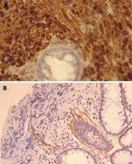
Figure 2 Immunohistochemical staining.
The lesion consists of a pure population of Schwann cells, as shown by the diffuse immunoreactivity for the S-100 protein (A). Only scattered myoepithelial cells and vascular structures were highlighted by the immunostaining for α-smooth muscle actin (B).
- Citation: Pasquini P, Baiocchini A, Falasca L, Annibali D, Gimbo G, Pace F, Nonno FD. Mucosal Schwann cell “Hamartoma”: A new entity? World J Gastroenterol 2009; 15(18): 2287-2289
- URL: https://www.wjgnet.com/1007-9327/full/v15/i18/2287.htm
- DOI: https://dx.doi.org/10.3748/wjg.15.2287


