Copyright
©2008 The WJG Press and Baishideng.
World J Gastroenterol. Mar 7, 2008; 14(9): 1450-1452
Published online Mar 7, 2008. doi: 10.3748/wjg.14.1450
Published online Mar 7, 2008. doi: 10.3748/wjg.14.1450
Figure 1 CT scan on admission.
A: Mid-sagital view: Air in posterior retropharyngeal space, Bone fragment (*); B: Axial view at the level of C7. Bone fragment and esophageal perforation (*); C: Axial view at the level of C4 (hyoid bone): Air in posterior retropharyngeal space; D: Axial view at the level of T3: Pleural effusion and air bubbles in postero-superior mediastinum → Oe: esophagus.
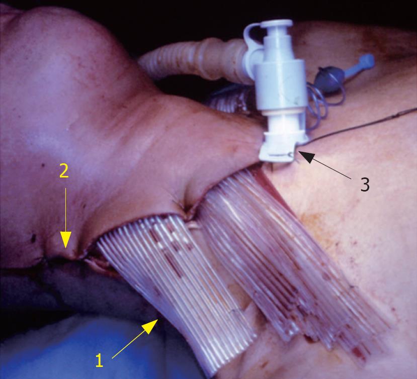
Figure 2 Surgical site at the end of the procedure.
1: Multitubular silicone drain; 2:
Neck incision; 3: Tracheotomy tube.
- Citation: Righini CA, Tea BZ, Reyt E, Chahine KA. Cervical cellulitis and mediastinitis following esophageal perforation: A case report. World J Gastroenterol 2008; 14(9): 1450-1452
- URL: https://www.wjgnet.com/1007-9327/full/v14/i9/1450.htm
- DOI: https://dx.doi.org/10.3748/wjg.14.1450


