Copyright
©2008 The WJG Press and Baishideng.
World J Gastroenterol. Aug 14, 2008; 14(30): 4841-4843
Published online Aug 14, 2008. doi: 10.3748/wjg.14.4841
Published online Aug 14, 2008. doi: 10.3748/wjg.14.4841
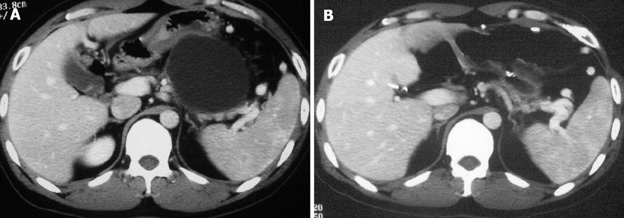
Figure 1 A: Pre-operative CT scan shows a 7.
5 cm X 6.0 cm pseudocyst, is in close contact with the posterior wall of the stomach. In addition, splenic vein compression and splenomegaly are also observed. B: Post-operative CT scan reveals effective drainage of the pseudocyst and dramatic relief of splenic vein compression.
- Citation: Sheng QS, Chen DZ, Lang R, Jin ZK, Han DD, Li LX, Yang YJ, Li P, Pan F, Zhang D, Qu ZW, He Q. Laparoscopic cystogastrostomy for the treatment of pancreatic pseudocysts: A case report. World J Gastroenterol 2008; 14(30): 4841-4843
- URL: https://www.wjgnet.com/1007-9327/full/v14/i30/4841.htm
- DOI: https://dx.doi.org/10.3748/wjg.14.4841


