Copyright
©2008 The WJG Press and Baishideng.
World J Gastroenterol. Aug 14, 2008; 14(30): 4826-4829
Published online Aug 14, 2008. doi: 10.3748/wjg.14.4826
Published online Aug 14, 2008. doi: 10.3748/wjg.14.4826
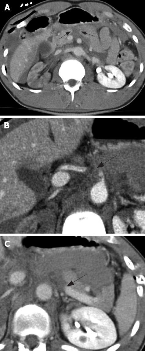
Figure 1 CT scan shows pancreatic transection between the head and body (A), thrombosis of the hepatic artery at its origin from the celiac trunk (B) and rupture/thrombosis of the splenic vein (C).
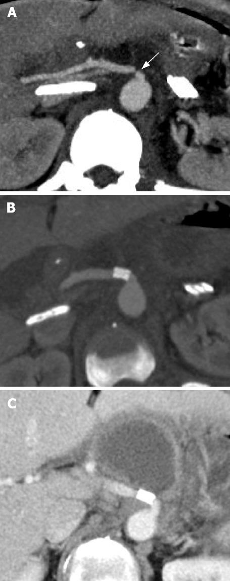
Figure 2 CT scan 12 h after operation showing severe stenosis of hepatic artery-celiac reconstruction (A), good result of the procedure confirmed by CT after endovascular treatment (B), and a symptomatic pseudocyst in the epigastric region three months later (C) which was successfully treated by percutaneous drainage, rest and antibiotics.
- Citation: Baiocchi GL, Tiberio GA, Gheza F, Gardani M, Cantù M, Portolani N, Giulini SM. Pancreatic transection from blunt trauma associated with vascular and biliary lesions: A case report. World J Gastroenterol 2008; 14(30): 4826-4829
- URL: https://www.wjgnet.com/1007-9327/full/v14/i30/4826.htm
- DOI: https://dx.doi.org/10.3748/wjg.14.4826


