Copyright
©2008 The WJG Press and Baishideng.
World J Gastroenterol. Aug 14, 2008; 14(30): 4823-4825
Published online Aug 14, 2008. doi: 10.3748/wjg.14.4823
Published online Aug 14, 2008. doi: 10.3748/wjg.14.4823
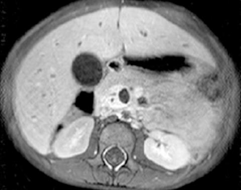
Figure 1 Contrast enhanced abdominal MRI shows a crescent shaped enhancing soft-tissue density within a dilated CBD.
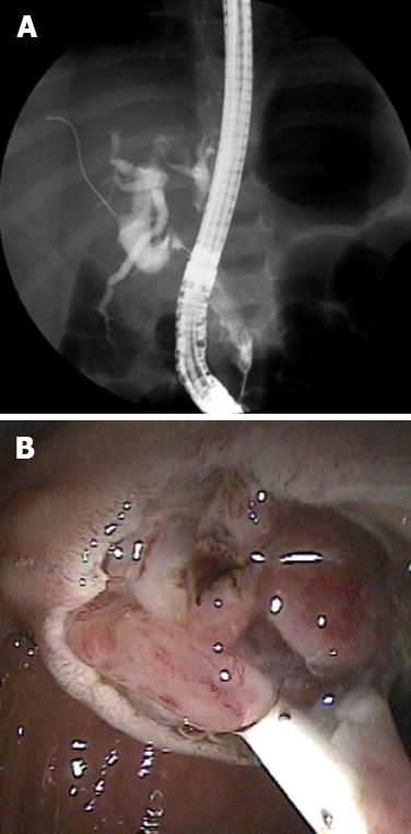
Figure 2 A: ERCP demonstrates filling defects along the dilated CBD and a diffusely dilated intrahepatic and extrahepatic biliary tree; B: Intraductal tumor protruding from the ampulla post-sphincterotomy.
A 7-french biliary stent was placed for decompression.
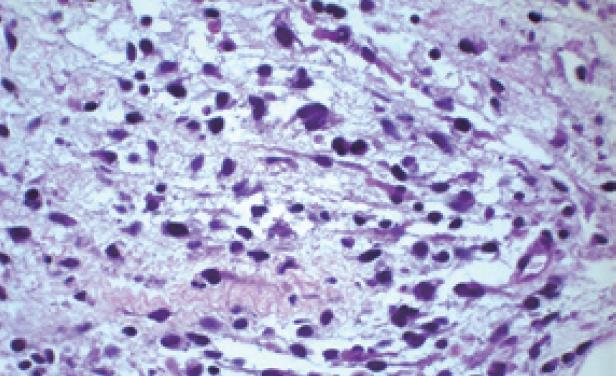
Figure 3 A few rhabdomyosarcoma cells have eosinophilic cytoplasm suggesting skeletal muscle differentiation but most are very undifferentiated in a myxoid stroma (HE, × 400).
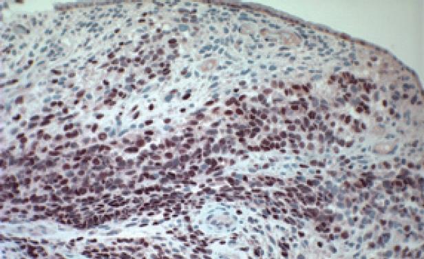
Figure 4 Immunostain for myogenin reveals almost 100% positivity in neoplastic cell nuclei.
The growth of this tumor beneath biliary epithelium (top) is typical of botryoid rhabdomyosarcoma (× 250).
- Citation: Himes RW, Raijman I, Finegold MJ, Russell HV, Fishman DS. Diagnostic and therapeutic role of endoscopic retrograde cholangiopancreatography in biliary rhabdomyosarcoma. World J Gastroenterol 2008; 14(30): 4823-4825
- URL: https://www.wjgnet.com/1007-9327/full/v14/i30/4823.htm
- DOI: https://dx.doi.org/10.3748/wjg.14.4823


