Copyright
©2008 The WJG Press and Baishideng.
World J Gastroenterol. Aug 14, 2008; 14(30): 4816-4822
Published online Aug 14, 2008. doi: 10.3748/wjg.14.4816
Published online Aug 14, 2008. doi: 10.3748/wjg.14.4816
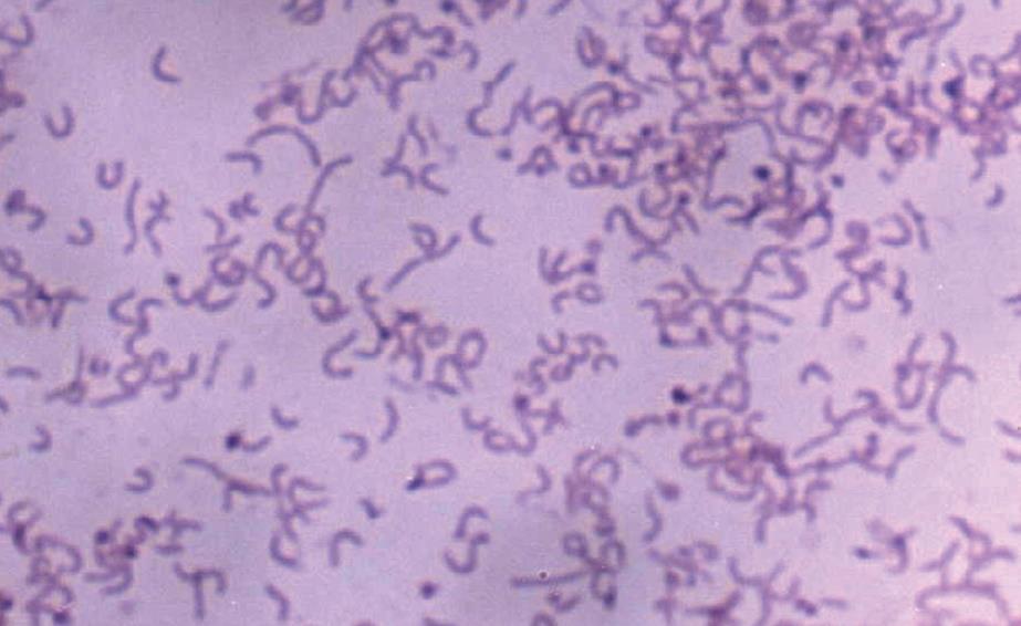
Figure 1 H pylori isolated from a patients’ biopsy (Gram-staining, × 1000).
Figure 2 Expression and purification of target recombinant proteins.
M: Marker (BioColor); Lane 1: Blank expression vector; Lanes 2-8 and 9-15: rUreB, rHpaA, rVacA, rCagA1, rNapA, rFlaA and rFlaB, respectively.
Figure 3 Detection results of target recombinant proteins by Western blot assay.
M: Marker (BioColor); Lanes 1a-7a: rUreB, rHpaA, rVacA, rCagA1, rNapA, rFlaA, rFlaB, respectively; Lanes 1b-7b: blank controls.
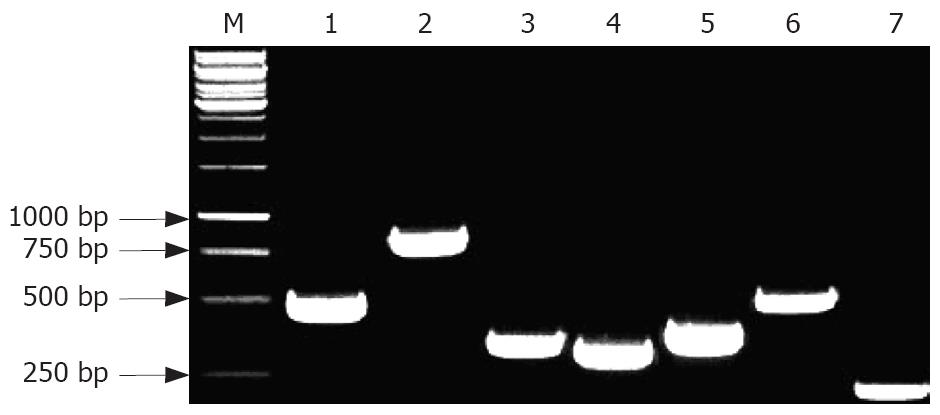
Figure 4 PCR products of 7 genes of H pylori.
M: Marker (BioColor); Lanes 1-7: ureB, hpaA, vacA, CagA, napA, flaA and flab genes of H pylori NCTC 11637, respectively.
-
Citation: Tang RX, Luo DJ, Sun AH, Yan J. Diversity of
Helicobacter pylori isolates in expression of antigens and induction of antibodies. World J Gastroenterol 2008; 14(30): 4816-4822 - URL: https://www.wjgnet.com/1007-9327/full/v14/i30/4816.htm
- DOI: https://dx.doi.org/10.3748/wjg.14.4816


