Copyright
©2008 The WJG Press and Baishideng.
World J Gastroenterol. Jul 21, 2008; 14(27): 4407-4409
Published online Jul 21, 2008. doi: 10.3748/wjg.14.4407
Published online Jul 21, 2008. doi: 10.3748/wjg.14.4407
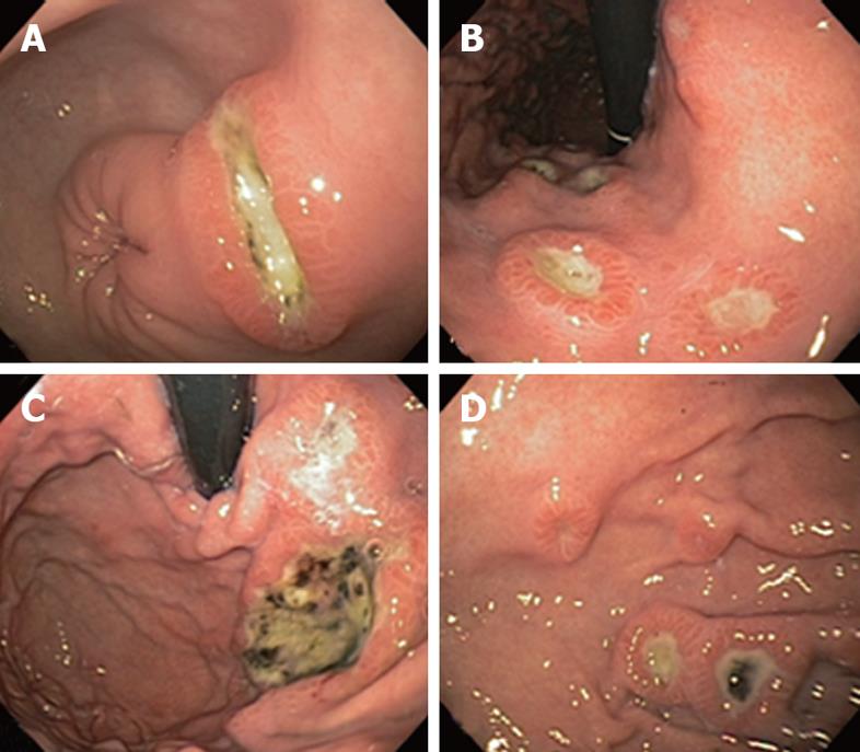
Figure 1 Endoscopic appearance of DLBCL infiltration of gastric antrum (A), lesser curvature (B), fundus (C), and body (D).
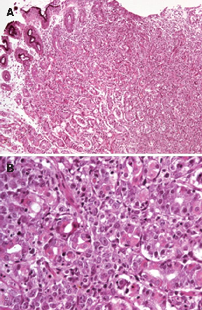
Figure 2 Gastric mucosa biopsy showing diffuse infiltration of lamina propria with distortion of the glandular architecture (A) and diffuse infiltration by large lymphoid cells (centroblast-type) that surround and destroy the gastric glands (B).
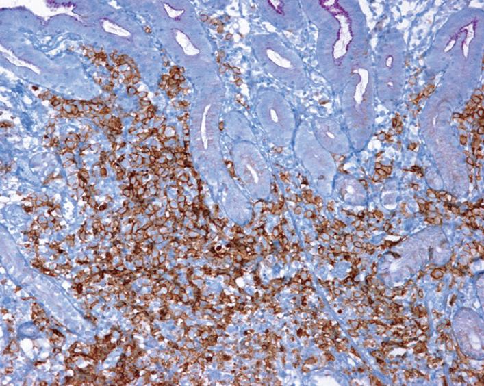
Figure 3 Gastric biopsy showing infiltration by atypical lymphoid cells in the gastric mucosa with intense positivity for CD20 at immunohistochemical analysis.
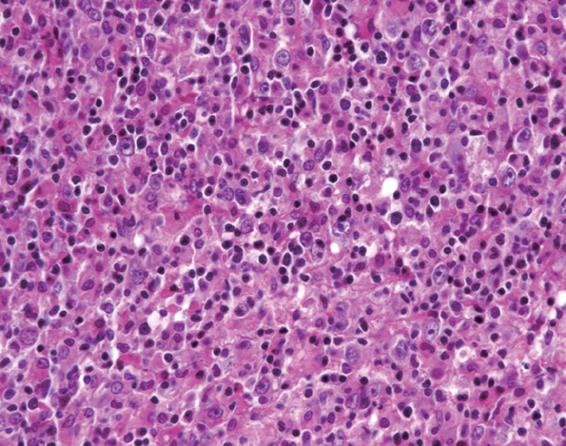
Figure 4 Lymph node biopsy showing neoplastic lymphocytes with fine nuclear chromatin, some of which have 2 or 3 peripheral nucleoli (centroblastic type) and others have prominent central nucleoli (immunoblastic type).
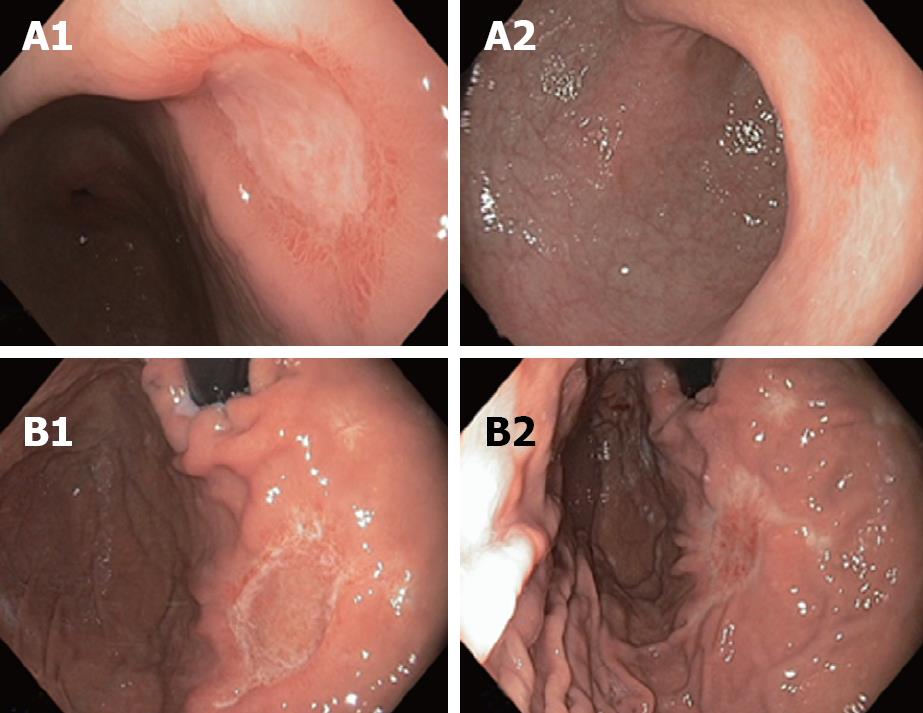
Figure 5 Endoscopic appearances of gastric antrum (A1, A2) and fundus (B1, B2) after 3 and 6 cycles of chemotherapy respectively.
- Citation: Zepeda-Gómez S, Camacho J, Oviedo-Cárdenas E, Lome-Maldonado C. Gastric infiltration of diffuse large B-cell lymphoma: Endoscopic diagnosis and improvement of lesions after chemotherapy. World J Gastroenterol 2008; 14(27): 4407-4409
- URL: https://www.wjgnet.com/1007-9327/full/v14/i27/4407.htm
- DOI: https://dx.doi.org/10.3748/wjg.14.4407


